Effects of treadmill running and limb immobilization on knee cartilage degeneration and locomotor joint kinematics in rats following knee meniscal transection
- PMID: 31437580
- PMCID: PMC7576441
- DOI: 10.1016/j.joca.2019.08.001
Effects of treadmill running and limb immobilization on knee cartilage degeneration and locomotor joint kinematics in rats following knee meniscal transection
Abstract
Objective: This study examined the effects of reduced and elevated weight bearing on post-traumatic osteoarthritis (PTOA) development, locomotor joint kinematics, and degree of voluntary activity in rats following medial meniscal transection (MMT).
Design: Twenty-one adult rats were subjected to MMT surgery of the left hindlimb and then assigned to one of three groups: (1) regular (i.e., no intervention), (2) hindlimb immobilization, or (3) treadmill running. Sham surgery was performed in four additional rats. Voluntary wheel run time/distance was measured, and 3D hindlimb kinematics were quantified during treadmill locomotion using biplanar radiography. Rats were euthanized 8 weeks after MMT or sham surgery, and the microstructure of the tibial cartilage and subchondral bone was quantified using contrast enhanced micro-CT.
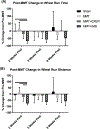
Results: All three MMT groups showed signs of PTOA (full-thickness lesions and/or increased cartilage volume) compared to the sham group, however the regular and treadmill-running groups had greater osteophyte formation than the immobilization group. For the immobilization group, increased volume was only observed in the anterior region of the cartilage. The treadmill-running group demonstrated a greater knee varus angle at mid-stance than the sham group, while the immobilization group demonstrated greater reduction in voluntary running than all the other groups at 2 weeks post-surgery.
Conclusions: Elevated weight-bearing via treadmill running at a slow/moderate speed did not accelerate PTOA in MMT rats when compared to regular weight-bearing. Reduced weight-bearing via immobilization may attenuate overall PTOA but still resulted in regional cartilage degeneration. Overall, there were minimal differences in hindlimb kinematics and voluntary running between MMT and sham rats.
Keywords: Computed tomography; Knee injury; Osteoarthritis; Rehabilitation; X-ray motion analysis.
Copyright © 2019 Osteoarthritis Research Society International. Published by Elsevier Ltd. All rights reserved.
Conflict of interest statement
Conflict of interests
All authors have no conflict of interests to declare.
Figures


Similar articles
-
Pain and knee damage in male and female mice in the medial meniscal transection-induced osteoarthritis.Osteoarthritis Cartilage. 2020 Apr;28(4):475-485. doi: 10.1016/j.joca.2019.11.003. Epub 2019 Dec 10. Osteoarthritis Cartilage. 2020. PMID: 31830592
-
Physiological exercise loading suppresses post-traumatic osteoarthritis progression via an increase in bone morphogenetic proteins expression in an experimental rat knee model.Osteoarthritis Cartilage. 2017 Jun;25(6):964-975. doi: 10.1016/j.joca.2016.12.008. Epub 2016 Dec 10. Osteoarthritis Cartilage. 2017. PMID: 27965139
-
Accelerated post traumatic osteoarthritis in a dual injury murine model.Osteoarthritis Cartilage. 2019 Dec;27(12):1800-1810. doi: 10.1016/j.joca.2019.05.027. Epub 2019 Jul 5. Osteoarthritis Cartilage. 2019. PMID: 31283983
-
Subchondral plate porosity colocalizes with the point of mechanical load during ambulation in a rat knee model of post-traumatic osteoarthritis.Osteoarthritis Cartilage. 2016 Feb;24(2):354-63. doi: 10.1016/j.joca.2015.09.001. Epub 2015 Sep 14. Osteoarthritis Cartilage. 2016. PMID: 26376125
-
The effect of anti-gravity training after meniscal or chondral injury in the knee. A systematic review.Acta Orthop Belg. 2020 Sep;86(3):422-433. Acta Orthop Belg. 2020. PMID: 33581026
Cited by
-
Wheel Running Exacerbates Joint Damage after Meniscal Injury in Mice, but Does Not Alter Gait or Physical Activity Levels.Med Sci Sports Exerc. 2023 Sep 1;55(9):1564-1576. doi: 10.1249/MSS.0000000000003198. Epub 2023 May 5. Med Sci Sports Exerc. 2023. PMID: 37144624 Free PMC article.
-
Teaming up to overcome challenges toward translation of new therapeutics for osteoarthritis.J Orthop Res. 2024 Dec;42(12):2659-2672. doi: 10.1002/jor.25944. Epub 2024 Aug 5. J Orthop Res. 2024. PMID: 39103981
-
37-Day microgravity exposure in 16-Week female C57BL/6J mice is associated with bone loss specific to weight-bearing skeletal sites.PLoS One. 2025 Mar 26;20(3):e0317307. doi: 10.1371/journal.pone.0317307. eCollection 2025. PLoS One. 2025. PMID: 40138271 Free PMC article.
-
Articular Cartilage- and Synoviocyte-Binding Poly(ethylene glycol) Nanocomposite Microgels as Intra-Articular Drug Delivery Vehicles for the Treatment of Osteoarthritis.ACS Biomater Sci Eng. 2020 Sep 14;6(9):5084-5095. doi: 10.1021/acsbiomaterials.0c00960. Epub 2020 Aug 28. ACS Biomater Sci Eng. 2020. PMID: 33455260 Free PMC article.
-
Mechanical loading and orthobiologic therapies in the treatment of post-traumatic osteoarthritis (PTOA): a comprehensive review.Front Bioeng Biotechnol. 2024 Jun 24;12:1401207. doi: 10.3389/fbioe.2024.1401207. eCollection 2024. Front Bioeng Biotechnol. 2024. PMID: 38978717 Free PMC article. Review.
References
-
- Meunier A, Odensten M, Good L. Long-term results after primary repair or non-surgical treatment of anterior cruciate ligament rupture: a randomized study with a 15-year follow-up. Scand J Med Sci Sport 2007;17:230–7. - PubMed
-
- Tammi M, Kiviranta I, Peltonen L, Jurvelin J, Helminen HJ. Effects of joint loading on articular cartilage collagen metabolism: assay of procollagen prolyl 4-hydroxylase and galactosylhydroxylysyl glucosyltransferase. Connect Tissue Res 1988;17:199–206. - PubMed
Publication types
MeSH terms
Grants and funding
LinkOut - more resources
Full Text Sources

