

Case report: a peculiar glomerulopathy in a patient suffering from nephrotic syndrome
- PMID: 31438874
- PMCID: PMC6704495
- DOI: 10.1186/s12882-019-1478-8
Case report: a peculiar glomerulopathy in a patient suffering from nephrotic syndrome
Abstract
Background: Podocyte infolding glomerulopathy (PIG) is a rare histopathologic finding with global infolding of the podocytes into the glomerular basement membrane (GBM), accompanied by microstructures underneath. Described in 2002 for the first time, PIG was proposed as a new pathological entity in 2008 based on the largest case series so far. Yet all of the described cases derive from Asian countries. We report a case from Germany fulfilling the diagnostic criteria of PIG. Considering the scarcity of data on this entity especially in Western countries, collecting cases like ours and multicentric meta-analyses will be crucial to obtain a better understanding of PIG, its causes, clinical course and potential treatment options.
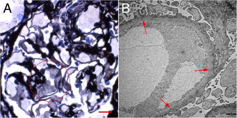
Case presentation: A 56-year-old Caucasian woman with a history of rheumatoid arthritis (RA), no other comorbidities and no known renal disease was admitted to the hospital with acute kidney injury (AKI) and nephrotic syndrome. Physical examination was unremarkable except for anasarca. Renal ultrasound revealed no abnormalities. Laboratory and urine analyses were consistent with the nephrotic syndrome and renal failure. Serological studies regarding ANA, ANCA, anti-PLA2R autoantibodies, complement, virus infections, immunofixation and quantitative light chain analysis were unremarkable. A renal biopsy was performed. Light microscopic examination showed flattened tubular epithelium consistent with acute tubular damage, no infiltrates and unremarkable glomeruli except diffuse and global holes in the GBM (Fig. 1a) and negative staining for immunoglobulin heavy-chains, light-chains and complement split products. Electron microscopy revealed a rare correlate for these holes: global peculiar infolding of podocyte cytoplasm into the GBM. Most of these infoldings were accompanied by condensation of the GBM underneath. No such condensation or electron dense deposits were found without these infoldings or outside the GBM.
Conclusion: Here we report the first case of PIG outside of Asia. Since there are only few reports about this specific finding, we feel there is a need to share information in an attempt to accumulate knowledge about this possible new entity and potential treatment options.
Keywords: Membranous Glomerulopathy; Microspheres; Nephrotic syndrome; Podocyte infolding; Renal biopsy.
Conflict of interest statement
The authors declare no conflict of interest. The results presented in this paper have not been published previously in whole or part anywhere else.
Figures
References
-
- Joh K, Taguchi T, Shigematsu H, Kobayashi Y, Sato H, Nishi S, Katafuchi R, Nomura S, Fujigaki Y, Utsunomiya Y, Sugiyama H, Saito T, Makino H. Proposal of podocytic infolding glomerulopathy as a new disease entity. A review of 25 cases from nationwide research in Japan. Clin Exp Nephrol. 2008;12:421–431. doi: 10.1007/s10157-008-0104-z. - DOI - PubMed
Publication types
MeSH terms
LinkOut - more resources
Full Text Sources
