

Reduction of metastatic potential by inhibiting EGFR/Akt/p38/ERK signaling pathway and epithelial-mesenchymal transition after carbon ion exposure is potentiated by PARP-1 inhibition in non-small-cell lung cancer
- PMID: 31438892
- PMCID: PMC6704719
- DOI: 10.1186/s12885-019-6015-4
Reduction of metastatic potential by inhibiting EGFR/Akt/p38/ERK signaling pathway and epithelial-mesenchymal transition after carbon ion exposure is potentiated by PARP-1 inhibition in non-small-cell lung cancer
Abstract
Background: Carbon ion (12C) radiotherapy is becoming very promising to kill highly metastatic cancer cells keeping adjacent normal cells least affected. Our previous study shows that combined PARP-1 inhibition with 12C ion reduces MMP-2,-9 synergistically in HeLa cells but detailed mechanism are not clear. To understand this mechanism and the rationale of using PARP-1 inhibitor with 12C ion radiotherapy for better outcome in controlling metastasis, we investigated metastatic potential in two non-small cell lung cancer (NSCLC) A549 and H1299 (p53-deficient) cells exposed with 12C ion in presence and absence of PARP-1 inhibition using siRNA or olaparib.
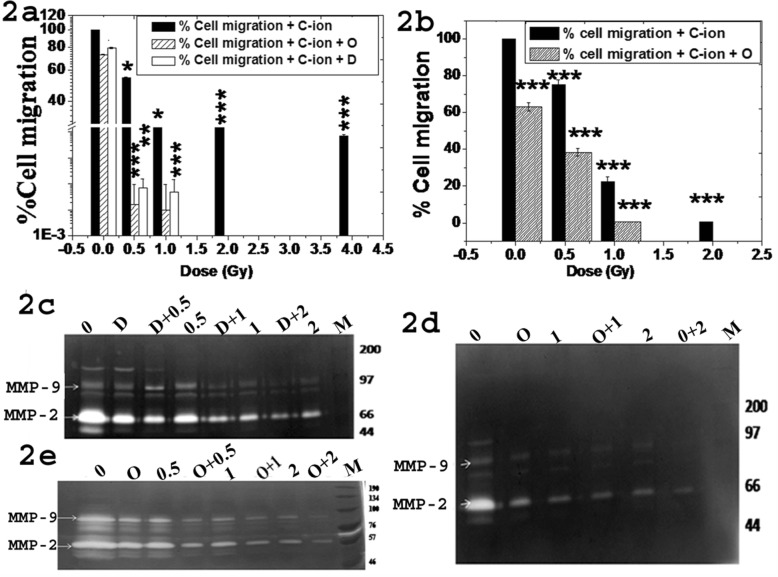
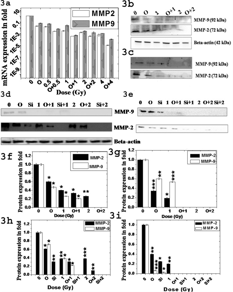
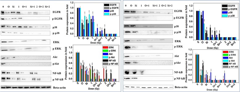
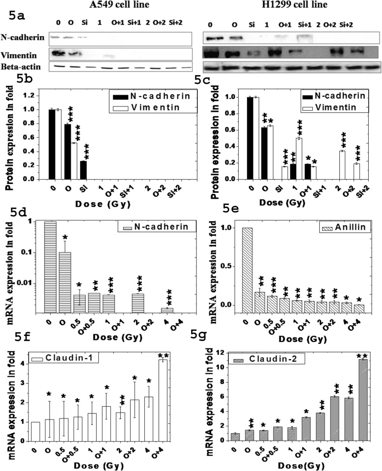
Methods: We monitored cell proliferation, in-vitro cell migration, wound healing, expression and activity of MMP-2, - 9 in A549 and p53-deficient H1299 cell lines exposed with 12C ion with and without PARP-1 inhibitor olaparib/DPQ. Expression and phosphorylation of NF-kB, EGFR, Akt, p38, ERK was also observed in A549 and H1299 cells exposed with 12C ion with and without PARP-1 inhibition using siRNA or olaparib. We also checked expression of few marker genes involved in epithelial-mesenchymal transition (EMT) pathways like N-cadherin, vimentin, anillin, claudin-1, - 2 in both NSCLC. To determine the generalized effect of 12C ion and olaparib in inhibition of cell's metastatic potential, wound healing and activity of MMP-2, - 9 was also studied in HeLa and MCF7 cell lines after 12C ion exposure and in combination with PARP-1 inhibitor olaparib.
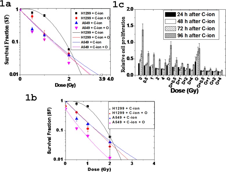
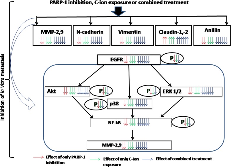
Results: Our experiments show that 12C ion and PARP-1 inhibition separately reduces cell proliferation, cell migration, wound healing, phosphorylation of EGFR, Akt, p38, ERK resulting inactivation of NF-kB. Combined treatment abolishes NF-kB expression and hence synergistically reduces MMP-2, - 9 expressions. Each single treatment reduces N-cadherin, vimentin, anillin but increases claudin-1, - 2 leading to suppression of EMT process. However, combined treatment synergistically alters these proteins to suppress EMT pathways significantly.
Conclusion: The activation pathways of transcription of MMP-2,-9 via NF-kB and key marker proteins in EMT pathways are targeted by both 12C ion and olaparib/siRNA. Hence, 12C ion radiotherapy could potentially be combined with olaparib as chemotherapeutic agent for better control of cancer metastasis.
Keywords: Carbon ion exposure; Epithelial-mesenchymal transition (EMT); Matrix metalloproteinases (MMPs); Metastatic potential; Non-small-cell lung cancer; PARP-1.
Conflict of interest statement
The authors declare that they have no competing interests.
Figures
References
-
- Ferlay J, Soerjomataram I, Dikshit R, Eser S, Mathers C, Rebelo M, et al. Cancer incidence and mortality worldwide: sources, methods and major patterns in GLOBOCAN 2012. Int J Cancer. 2015;136(5):E359–E386. - PubMed
-
- Siegel R, Ma J, Zou Z, Jemal A. Cancer statistics, 2014. CA Cancer J Clin. 2014;64(1):9–29 Available from: http://onlinelibrary.wiley.com/doi/10.3322/caac.21208/full%5Cnhttps://www.ncbi.nlm.nih.gov/pubmed/24399786. - DOI - PubMed
-
- Zhou YC, Liu JY, Li J, Zhang J, Xu YQ, Zhang HW, et al. Ionizing radiation promotes migration and invasion of cancer cells through transforming growth factor-beta-mediated epithelial-mesenchymal transition. Int J Radiat Oncol Biol Phys. 2011;81(5):1530–1537. - PubMed
-
- Loeffier JS, Durante M. Charged particle therapy- optimization, challenges and future directions. Nat Rev Clin Oncol. 2013;10:411–424. - PubMed
MeSH terms
Substances
Grants and funding
LinkOut - more resources
Full Text Sources
Medical
Research Materials
Miscellaneous
