. 2019 Aug 22;18(1):127.
doi: 10.1186/s12943-019-1053-8.

WTAP facilitates progression of hepatocellular carcinoma via m6A-HuR-dependent epigenetic silencing of ETS1

Affiliations

WTAP facilitates progression of hepatocellular carcinoma via m6A-HuR-dependent epigenetic silencing of ETS1

Yunhao Chen et al. Mol Cancer. .

Abstract

Background: N6-methyladenosine (m6A) methylation, a well-known modification with new epigenetic functions, has been reported to participate in the tumorigenesis of hepatocellular carcinoma (HCC), providing novel insights into the molecular pathogenesis of this disease. However, as the key component of m6A methylation, Wilms tumor 1-associated protein (WTAP) has not been well studied in HCC. Here we investigated the biological role and underlying mechanism of WTAP in liver cancer.

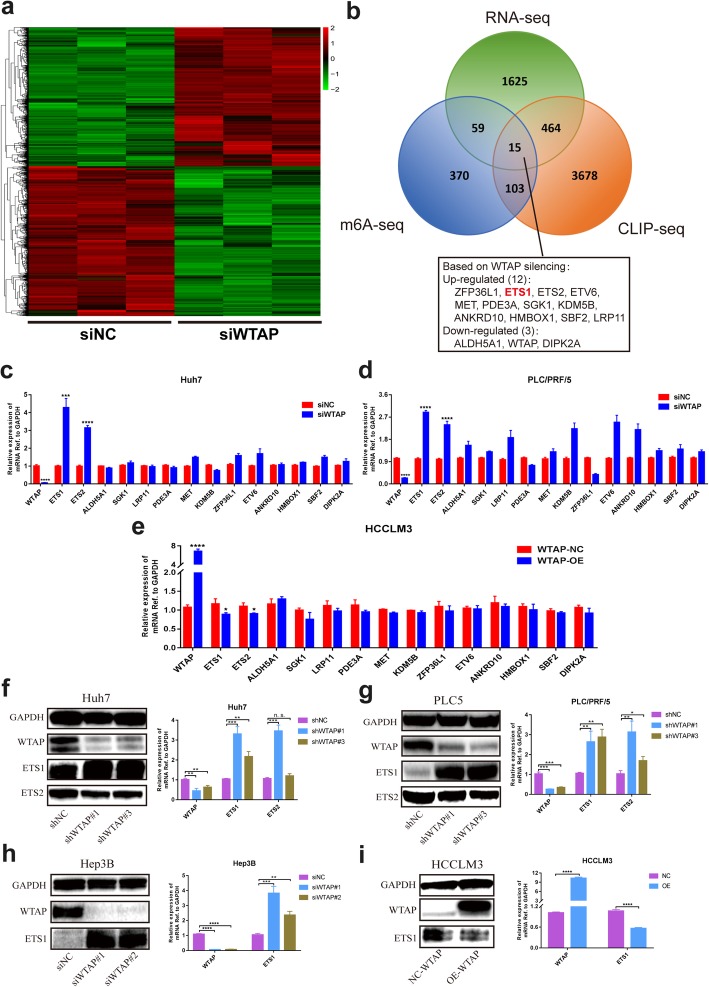
Methods: We determined the expression of WTAP and its correlation with clinicopathological features using tissue microarrays and the Cancer Genome Atlas (TCGA) dataset. And we clarified the effects of WTAP on HCC cells using cell proliferation assay, colony formation, Edu assay and subcutaneous xenograft experiments. We then applied RNA sequencing combined with gene expression omnibus (GEO) data to screen candidate targets of WTAP. Finally, we investigated the regulatory mechanism of WTAP in HCC by m6A dot blot assay, methylated RNA immunoprecipitation (MeRIP) assay, dual luciferase reporter assay, RNA immunoprecipitation (RIP) assay and Chromatin immunoprecipitation (ChIP) assay.

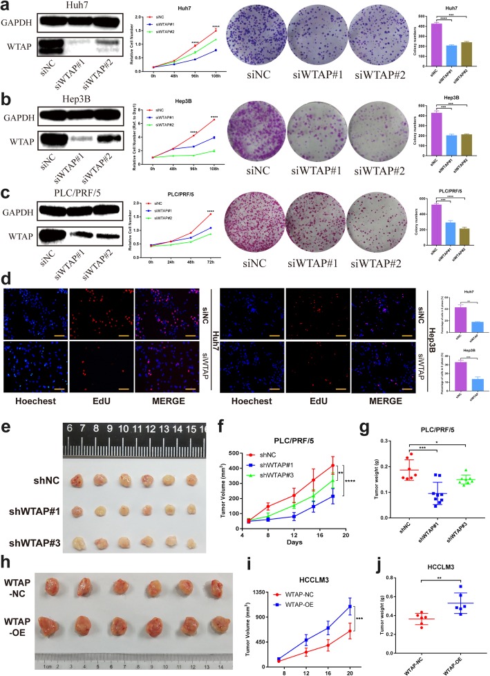
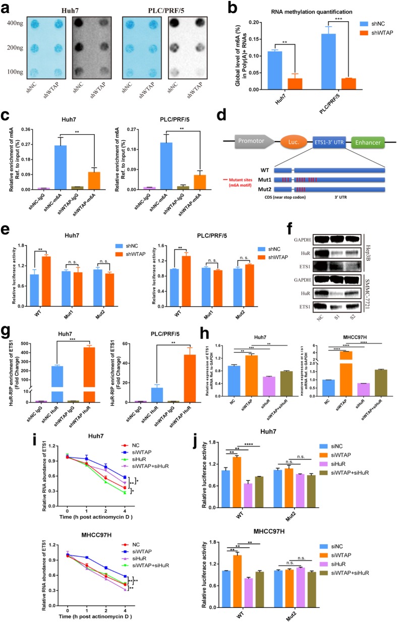
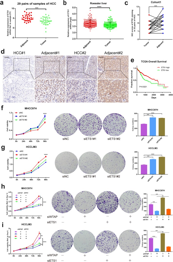
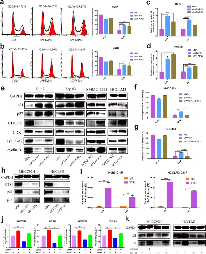
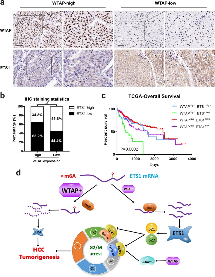
Results: We demonstrated that WTAP was highly expressed in HCC which indicated the poor prognosis, and that WTAP expression served as an independent predictor of HCC survival. Functionally, WTAP promoted the proliferation capability and tumor growth of HCC cells in vitro and in vivo. Furthermore, ETS proto-oncogene 1 (ETS1) was identified as the downstream effector of WTAP. The m6A modification regulated by WTAP led to post-transcriptional suppression of ETS1, with the implication of Hu-Antigen R (HuR) as an RNA stabilizer. Then ETS1 was found to inhibit the progression of HCC and could rescue the phenotype induced by WTAP deficiency. Moreover, WTAP modulated the G2/M phase of HCC cells through a p21/p27-dependent pattern mediated by ETS1.

Conclusion: We have identified that WTAP is significantly up-regulated in HCC and promotes liver cancer development. WTAP-guided m6A modification contributes to the progression of HCC via the HuR-ETS1-p21/p27 axis. Our study is the first to report that WTAP-mediated m6A methylation has a crucial role in HCC oncogenesis, and highlights WTAP as a potential therapeutic target of HCC treatment.

Keywords: ETS1; Hepatocellular carcinoma (HCC); N6-methyladenosine (m6A); Wilms tumor 1-associated protein (WTAP).


Conflict of interest statement

The authors declare that they have no competing interests.

Figures

Fig. 1
Fig. 1
Up-regulated WTAP expression is associated with poor outcomes of HCC. a The expression of WTAP mRNA was determined based on GEO datasets (GSE14520); b The expression of WTAP protein was analyzed by western blotting in 15 pairs of HCC tissues; (T: tumor; P: peritumor); c Representative IHC images of WTAP staining in HCC tumor or adjacent tissues (scale bar, 100 μm; magnification, 200X and 400X); d IHC scores of 90 pairs of HCC tissues in the TMA cohort based on WTAP staining; e Kaplan-Meier analysis of overall survival and disease free survival of 90 HCC patients (data from TMA); f Forest plots based upon the outcomes of multivariate analysis of several factors associated with OS and RFS of HCC patients. Note: The factors that were closely associated with clinical outcomes of HCC were adopted into a COX regression model. Therefore, the restriction of statistical significance (P < 0.05) may be properly broadened
Fig. 2
Fig. 2
WTAP promotes tumor growth of HCC cells in vitro and in vivo. a, b and c Negative control or siRNA (si-WTAP #1, #2) was transfected into Huh7 (a), Hep3B (b) and PLC/PRF/5 (c) cells, respectively. The knockdown efficiency was tested and proliferation capacities were detected by CCK-8 and colony formation assay, with bar charts showing colony numbers; d EdU assay was applied to compare the cell proliferation ability in Huh7 and Hep3B (scale bar, 100 μm); e, f and g Tumor growth curve (f) of stable WTAP silenced PLC/PRF/5 cells (or negative control) in the xenograft mouse model was based on tumor size measurement. Moreover, tumor nodules (e) were collected and tumor weights (g) were recorded to present the growth difference within the influence of WTAP. h, i and j Tumor growth curve (i) of stable WTAP overexpressing HCCLM3 cells (or negative control) in the xenograft model was presented, followed by the collection of tumor nodules (h) and tumor weight records (j)
Fig. 3
Fig. 3
A high-throughput sequencing combination revealed ETS1 to be the target of WTAP. a Transcriptome profiles from Huh7 cells transfected with the WTAP siRNAs or negative control siRNAs (both in triplicate) were shown. Bands with red, black or green in the heat map indicated high, moderate or low expression, respectively. b A Venn diagram was generated from the gene sets enriched for transcripts that were substantially altered after WTAP silencing (RNA-seq), along with those enriched for m6A-modified transcripts (m6A-seq) and those enriched for WTAP-conjugated transcripts (CLIP-seq). 15 genes were selected according to the overlaps. The RNA-seq data was acquired from our study, while the m6A-seq and CLIP-seq data were obtained from GEO datasets (GSE46705). Information regarding detailed gene sets of RNA-seq was listed in Additional file 4: Table S4; c, d and e RT-qPCR was performed in Huh7 (c) and PLC/PRF/5 (d) with WTAP silencing, and in HCCLM3 (e) with WTAP overexpression, to validate the overlapped genes. The variation of ETS1 and ETS2 was consistent among 15 genes in the above three cell lines; f and g Expression of ETS1 and ETS2 following WTAP knockdown was evaluated by western blotting and RT-qPCR in Huh7 (f) and PLC/PRF/5 cells (g); h and i Expression of ETS1 was further examined by western blotting and RT-qPCR in Hep3B (h) or HCCLM3 (i) cells following the knockdown or overexpression of WTAP
Fig. 4
Fig. 4
WTAP repressed ETS1 in an m6A-HuR mediated pattern. a The m6A level of poly(A) + RNAs isolated from total RNA of WTAP-knockdown Huh7 and PLC/PRF/5 cells was indicated by m6A dot blot. Corresponding RNAs were loaded equally by a 2-fold serial dilution with 400 ng, 200 ng and 100 ng. Methylene blue staining served as a loading control; b The global content of m6A was also examined by RNA methylation quantification assay, relying on the standard curve; c MeRIP analysis followed by qRT-PCR was applied to assess the m6A modification of ETS1 in two WTAP-silencing HCC cells. The enrichment of m6A in each group was calculated by m6A-IP/input and IgG-IP/input. d Three luciferase plasmids were constructed by inserting the corresponding cDNAs into pGL3-control vectors. Wild-type reporters embodied the full-length 3’UTR and a partial CDS sequence near stop codon of ETS1 with intact m6A sites, while mutant ones obtained some A-C mutations on m6A consensus motifs (Mut1 or Mut2 contained 11 or 4 mutations, respectively). Luciferase activity was detected and normalized to Renilla activity; e Relative activity of the WT or Mut luciferase reporters in WTAP-silenced Huh7 and PLC/PRF/5 cells was determined (normalized to negative control groups); f ETS1 expression was identified by western blotting in Hep3B and SMMC-7721 cells upon knockdown of HuR (#1, #2) compared with siNC; g Immunoprecipitation of HuR-related RNA in control or WTAP-knockdown cells was conducted followed by RT-qPCR to detect the amount of ETS1 mRNA binding to HuR; h ETS1 expression was measured by RT-qPCR in Huh7 and MHCC97H cells with or without knockdown of WTAP or HuR compared with NC; i The RNA decay rate was determined in Huh7 and MHCC97H cells after treatment with Actinomycin D (normalized to 0 h); j The relative activity of the WT or Mut luciferase reporters was detected in WTAP/HuR-rescued Huh7 and MHCC97H cells (normalized to negative control groups)
Fig. 5
Fig. 5
ETS1 played a tumor suppression role in HCC via the reversal of phenotypes mediated by WTAP. a Expression of ETS1 in HCC tumor and adjacent tissues from 29 pairs of HCC samples; b Expression of ETS1 mRNA was demonstrated based upon GEO datasets (GSE14520); c IHC scores of 32 pairs of HCC tissues in cohort1 based on ETS1 staining; d Representative IHC images of ETS1 staining in HCC tumor or adjacent tissues (scale bar, 100 μm; magnification, 200X and 400X); e Overall survival of HCC patients according to the level of ETS1 (data from TCGA); f and g CCK8 and colony formation assays were performed to examine the propagation ability of MHCC97H (f) and HCCLM3 (g), where ETS1 was knocked down or not, with bar charts indicating the colony numbers (right panel); h and i Rescue experiments were conducted to determine the influence of ETS1 silencing on WTAP knockdown cells (MHCC97H and HCCLM3), with bar charts showing colony numbers (right panel)
Fig. 6
Fig. 6
WTAP was involved in the cell cycle by alleviating the expression of p21 and p27. a and b Cell cycle distribution was analyzed by flow cytometry in Huh7 (a) and Hep3B (b) cells where WTAP were silenced, with summary bar charts showing the percentage of cells in each phase; c and d RT-qPCR was utilized to explore alterations of p21 and p27 when WTAP was knocked down in Huh7 (c) and Hep3B (d), respectively; e Cell cycle-related proteins including p21, p27, CDC25C, CDK1, cyclin-A2 and cyclin-B1 were measured by western blot in the indicated cells where WTAP was knocked down (Huh7 and Hep3B) or overexpressed (SMMC-7721 and HCCLM3); f and g Flow cytometry indicated that ETS1 knockdown could reverse the G2/M arrest in WTAP-silenced MHCC97H (f) or HCCLM3 (g) cell; h The knockdown efficiency of ETS1 was verified followed by the detection of p21 and p27 expression via western blotting; i The ChIP assay was conducted in Huh7 and HCCLM3 cells to determine whether ETS1 could bind to the promoter of p21 and p27 (IP/input was calculated); j and k A rescue assay was performed with or without knockdown of WTAP or ETS1 to validate the retrieved role of ETS1 in WTAP-mediated events. Expression of p21 and p27 were detected at the RNA (j) and protein (k) levels
Fig. 7
Fig. 7
High WTAP expression was correlated with low ETS1 expression and revealed a poor prognosis of HCC. a Representative IHC staining images of the identical HCC specimens with the staining of WTAP or ETS1 were shown, respectively (cohort1) (scale bar, 100 μm; magnification, 200X and 400X); b IHC results revealed that expression of WTAP and ETS1 was negatively interrelated in HCC tissues; c Overall survival analysis based on the co-expression of WTAP and ETS1 in HCC according to TCGA data; d A schematic model illustrating our findings on WTAP-mediated m6A regulation was shown

Similar articles

Cited by

References

    1. Siegel RL, Miller KD, Jemal A. Cancer statistics, 2017. CA Cancer J Clin. 2017;67:7–30. doi: 10.3322/caac.21387. - DOI - PubMed
    1. Ferlay J, Shin HR, Bray F, Forman D, Mathers C, Parkin DM. Estimates of worldwide burden of cancer in 2008: GLOBOCAN 2008. Int J Cancer. 2010;127:2893–2917. doi: 10.1002/ijc.25516. - DOI - PubMed
    1. El-Serag HB, Marrero JA, Rudolph L, Reddy KR. Diagnosis and treatment of hepatocellular carcinoma. Gastroenterology. 2008;134:1752–1763. doi: 10.1053/j.gastro.2008.02.090. - DOI - PubMed
    1. Villanueva A, Portela A, Sayols S, Battiston C, Hoshida Y, Mendez-Gonzalez J, Imbeaud S, Letouze E, Hernandez-Gea V, Cornella H, et al. DNA methylation-based prognosis and epidrivers in hepatocellular carcinoma. Hepatology. 2015;61:1945–1956. doi: 10.1002/hep.27732. - DOI - PubMed
    1. Desrosiers R, Friderici K, Rottman F. Identification of methylated nucleosides in messenger RNA from Novikoff hepatoma cells. Proc Natl Acad Sci U S A. 1974;71:3971–3975. doi: 10.1073/pnas.71.10.3971. - DOI - PMC - PubMed

Publication types

MeSH terms