Review
. 2019 Aug 22;12(1):84.
doi: 10.1186/s13045-019-0772-z.

Tumor-derived exosomes, myeloid-derived suppressor cells, and tumor microenvironment

Affiliations
Review

Tumor-derived exosomes, myeloid-derived suppressor cells, and tumor microenvironment

Xinyu Tian et al. J Hematol Oncol. .

Abstract

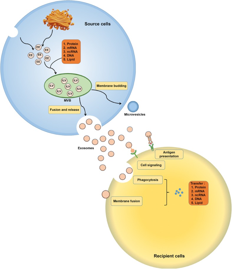
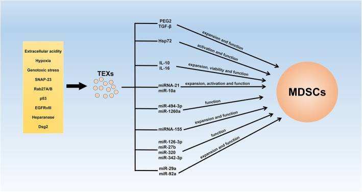
Plenty of immune cells infiltrate into the tumor microenvironment (TME) during tumor progression, in which myeloid-derived suppressor cells (MDSCs) represent a heterogeneous population of immature myeloid cells with immunosuppressive activity. Tumor cells and stromal cells facilitate the activation and expansion of MDSCs in TME via intercellular communication, and expanded MDSCs suppress anti-tumor immune responses through direct and indirect mechanisms. Currently, exosomes, which are a kind of extracellular vesicles (EVs) that can convey functional components, are demonstrated to participate in the local and distal intercellular communication between cells. Numerous studies have supposed that tumor-derived exosomes (TEXs), whose assembly and release can be modulated by TME, are capable of modulating the cell biology of MDSCs, including facilitating their activation, promoting the expansion, and enhancing the immunosuppressive function. Therefore, in this review, we mainly focus on the role of TEXs in the cell-cell communication between tumor cells and MDSCs, and discuss their clinical applications.

Keywords: Immunosuppression; Intercellular communication; Myeloid-derived suppressor cells; Tumor microenvironment; Tumor-derived exosomes.


Conflict of interest statement

The authors declare that they have no competing interests.

Figures

Fig. 1
Fig. 1
The formation and regulatory mechanism of exosomes. Exosome biogenesis initiates from the formation of EEs, which derive from the TGN and internalization of membrane microdomains. Then, EEs move into MVBs. During the inward budding of EEs into MVBs, vesicles load different cargoes and form ILVs. In this procedure, the loading of small plasma that contains nearly 100 proteins and 10000 nucleotides with proteins, coding and non-coding RNA, and DNA is a non-random process. Ras-related proteins regulate MVB movement towards cell membrane. MVBs fuse with the plasma membrane, and ILVs released to extracellular space are called exosomes. Exosomes received by recipient cells can be regarded as signalosomes for several biological processes. They can transfer both major histocompatibility complex (MHC) molecule and antigen, thereby involved in antigen presentation and immune regulation. Exosomes can also directly bind cell surface receptors and activate associated pathways. Additionally, exosomes can convey effectors including transcription factors, oncogenes, and infectious particles into recipient cells. Meanwhile, various nucleic acids are contained in extracellular vesicles and can be functionally delivered into recipient cells
Fig. 2
Fig. 2
TEXs participate in the cell-cell communication between tumor cells and MDSCs. Environmental conditions, such as extracellular acidity, hypoxia, genotoxic stress, and associated proteins in TME are capable of contributing to the formation and release of TEXs. Released TEXs are able to enhance the activation, expansion, and immunosuppression of MDSCs by conveying functional cargoes

References

    1. Becker A, Thakur BK, Weiss JM, Kim HS, Peinado H, Lyden D. Extracellular vesicles in cancer: cell-to-cell mediators of metastasis. Cancer Cell. 2016;30(6):836–848. doi: 10.1016/j.ccell.2016.10.009. - DOI - PMC - PubMed
    1. Choo YW, Kang M, Kim HY, Han J, Kang S, Lee JR, et al. M1 Macrophage-derived nanovesicles potentiate the anticancer efficacy of immune checkpoint inhibitors. ACS Nano. 2018;12(9):8977–8993. doi: 10.1021/acsnano.8b02446. - DOI - PubMed
    1. Wang Y, Tian J, Tang X, Rui K, Tian X, Ma J, et al. Exosomes released by granulocytic myeloid-derived suppressor cells attenuate DSS-induced colitis in mice. Oncotarget. 2016;7(13):15356–15368. - PMC - PubMed
    1. Robbins PD, Morelli AE. Regulation of immune responses by extracellular vesicles. Nat Rev Immunol. 2014;14(3):195–208. doi: 10.1038/nri3622. - DOI - PMC - PubMed
    1. Valenti R, Huber V, Filipazzi P, Pilla L, Sovena G, Villa A, et al. Human tumor-released microvesicles promote the differentiation of myeloid cells with transforming growth factor-beta-mediated suppressive activity on T lymphocytes. Cancer Res. 2006;66(18):9290–9298. doi: 10.1158/0008-5472.CAN-06-1819. - DOI - PubMed

Publication types

MeSH terms

LinkOut - more resources