. 2019:104:185-224.
doi: 10.1016/bs.aivir.2019.07.002. Epub 2019 Aug 7.

Hantavirus entry: Perspectives and recent advances

Affiliations

Hantavirus entry: Perspectives and recent advances

Eva Mittler et al. Adv Virus Res. 2019.

Abstract

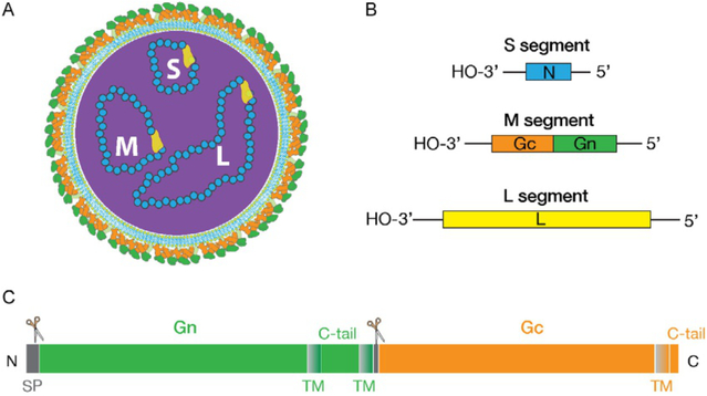
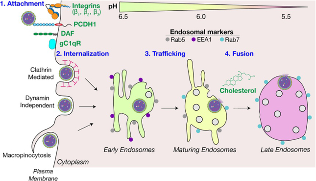
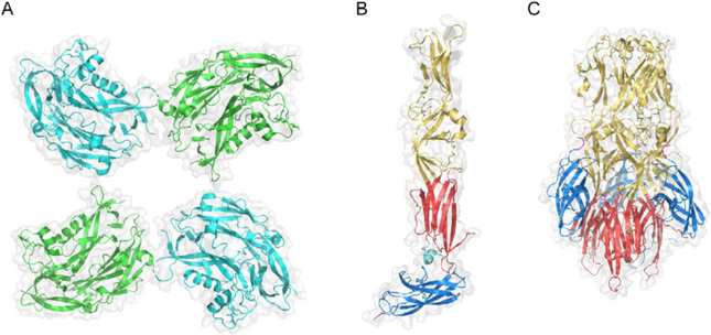
Hantaviruses are important zoonotic pathogens of public health importance that are found on all continents except Antarctica and are associated with hemorrhagic fever with renal syndrome (HFRS) in the Old World and hantavirus pulmonary syndrome (HPS) in the New World. Despite the significant disease burden they cause, no FDA-approved specific therapeutics or vaccines exist against these lethal viruses. The lack of available interventions is largely due to an incomplete understanding of hantavirus pathogenesis and molecular mechanisms of virus replication, including cellular entry. Hantavirus Gn/Gc glycoproteins are the only viral proteins exposed on the surface of virions and are necessary and sufficient to orchestrate virus attachment and entry. In vitro studies have implicated integrins (β1-3), DAF/CD55, and gC1qR as candidate receptors that mediate viral attachment for both Old World and New World hantaviruses. Recently, protocadherin-1 (PCDH1) was demonstrated as a requirement for cellular attachment and entry of New World hantaviruses in vitro and lethal HPS in vivo, making it the first clade-specific host factor to be identified. Attachment of hantavirus particles to cellular receptors induces their internalization by clathrin-mediated, dynamin-independent, or macropinocytosis-like mechanisms, followed by particle trafficking to an endosomal compartment where the fusion of viral and endosomal membranes can occur. Following membrane fusion, which requires cholesterol and acid pH, viral nucleocapsids escape into the cytoplasm and launch genome replication. In this review, we discuss the current mechanistic understanding of hantavirus entry, highlight gaps in our existing knowledge, and suggest areas for future inquiry.

Keywords: Bunyavirus; Hantavirus; Hantavirus pulmonary syndrome; Hemorrhagic fever with renal syndrome; Vaccines; Viral entry; Viral glycoprotein structure; Viral glycoproteins; Viral receptors.


Figures

Fig. 1
Fig. 1
Schematic representation of hantavirus particle and genes. (A) Hantaviruses carry a tri-segmented negative-sense RNA genome encased in a lipid envelope that is studded with surface spikes comprising the Gn and Gc glycoproteins. (B) Small (S), medium (M) and large (L) genome segments encode nucleoprotein (N), Gn/Gc glycoproteins and RNA-dependent RNA polymerase, respectively. (C) Linear representation of hantavirus Gn/Gc. Gn is proteolytically cleaved from Gc at the conserved WAASA site by the cellular signal protease complex (represented by scissors). SP, signal peptide; TM, transmembrane region (shown as shaded block); C-tail, cytoplasmic tail.
Fig. 2
Fig. 2
An overview of the hantavirus entry pathway. Host factors demonstrated or proposed to play a role in four stages of hantavirus entry, i.e., attachment, internalization, trafficking and membrane fusion are indicated. See the text for details.
Fig. 3
Fig. 3
Structure of hantavirus Gn and Gc. (A) Partial structural model for Gn tetramer derived by fitting the X-ray crystal structure of the N-terminal region of PUUV Gn (amino acid residues 29–383; PDB ID: 5FXU) into the cryo-electron tomography (cryo-ET)-derived density map of the TULV glycoprotein spike (PDB ID: 5FYN) (Li et al., 2016). Adjacent subunits are colored in cyan and green. Gray outlines in (A)–(C) represent surface-shaded views. En face view relative to the surface of the viral particle is shown. (B) X-ray crystal structure of the monomeric ectodomain of HTNV Gc in its putative pre-fusion conformation (PDB ID: 5LJY) (Guardado-Calvo et al., 2016). (C) X-ray crystal structure of the ectodomain of HTNV Gc in its trimeric post-fusion conformation (PDB ID: 5LK0) (Guardado-Calvo et al., 2016). (B) and (C) Gc is colored according to the convention for Class II fusion proteins: domains I, II, and III are shown in red, yellow, and blue, respectively. The DI–DIII linker is shown in cyan, and the beginning of the stem region at the C-terminus of the crystallized ectodomain constructs is shown in magenta. Disulfide bonds are shown as sticks, with sulfur atoms colored green. (C) The trimer is oriented axially to the viral and target membranes, with the fusion loops in domain II at the top of the structure.

References

    1. Acuña R, Cifuentes-Muñoz N, Márquez CL, Bulling M, Klingström J, Mancini R, Lozach P-Y, Tischler ND, 2014. Hantavirus Gn and Gc glycoproteins self-assemble into virus-like particles. J. Virol 88, 2344–2348. 10.1128/JVI.03118-13. - DOI - PMC - PubMed
    1. Acuña R, Bignon EA, Mancini R, Lozach P-Y, Tischler ND, 2015. Acidification triggers Andes hantavirus membrane fusion and rearrangement of Gc into a stable post-fusion homotrimer. J. Gen. Virol 96, 3192–3197. 10.1099/jgv.0.000269. - DOI - PubMed
    1. Albornoz A, Hoffmann AB, Lozach P-Y, Tischler ND, 2016. Early bunyavirus-host cell interactions. Viruses 8, 143 10.3390/v8050143. - DOI - PMC - PubMed
    1. Alff PJ, Sen N, Gorbunova E, Gavrilovskaya IN, Mackow ER, 2008. The NY-1 hantavirus Gn cytoplasmic tail coprecipitates TRAF3 and inhibits cellular interferon responses by disrupting TBK1-TRAF3 complex formation. J. Virol 82, 9115–9122. 10.1128/JVI.00290-08. - DOI - PMC - PubMed
    1. Aplin JD, Spanswick C, Behzad F, Kimber SJ, Vićovac L, 1996. Integrins beta 5, beta 3 and alpha v are apically distributed in endometrial epithelium. Mol. Hum. Reprod 2, 527–534. - PubMed

MeSH terms