Review
. 2019 Oct 11;294(41):15158-15171.
doi: 10.1074/jbc.REV119.006513. Epub 2019 Aug 22.

How do cells cope with RNA damage and its consequences?

Affiliations
Review

How do cells cope with RNA damage and its consequences?

Liewei L Yan et al. J Biol Chem. .

Abstract

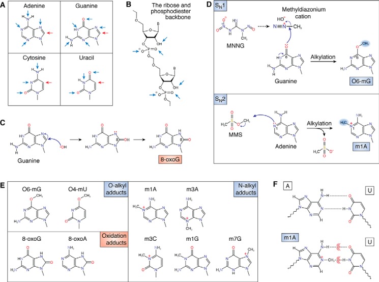
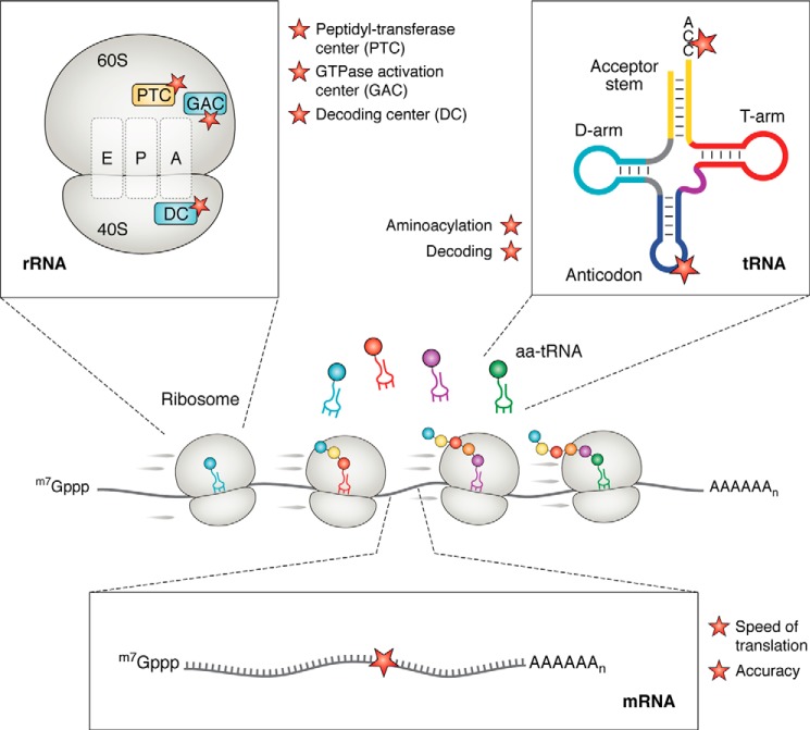
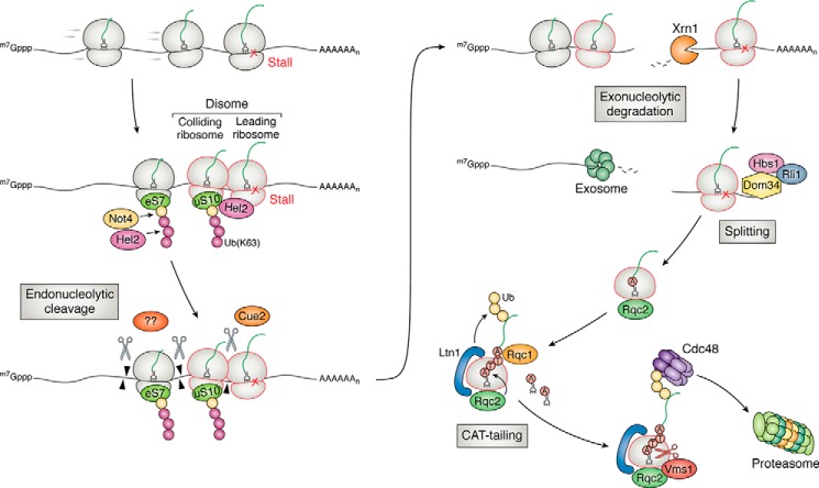
Similar to many other biological molecules, RNA is vulnerable to chemical insults from endogenous and exogenous sources. Noxious agents such as reactive oxygen species or alkylating chemicals have the potential to profoundly affect the chemical properties and hence the function of RNA molecules in the cell. Given the central role of RNA in many fundamental biological processes, including translation and splicing, changes to its chemical composition can have a detrimental impact on cellular fitness, with some evidence suggesting that RNA damage has roles in diseases such as neurodegenerative disorders. We are only just beginning to learn about how cells cope with RNA damage, with recent studies revealing the existence of quality-control processes that are capable of recognizing and degrading or repairing damaged RNA. Here, we begin by reviewing the most abundant types of chemical damage to RNA, including oxidation and alkylation. Focusing on mRNA damage, we then discuss how alterations to this species of RNA affect its function and how cells respond to these challenges to maintain proteostasis. Finally, we briefly discuss how chemical damage to noncoding RNAs such as rRNA, tRNA, small nuclear RNA, and small nucleolar RNA is likely to affect their function.

Keywords: Alzheimer disease; RNA; RNA damage; RNA modification; RNA repair; alkB; alkylation; mRNA surveillance; oxidative stress; quality control; ribosome; stress; translation; ubiquitin.


Conflict of interest statement

The authors declare that they have no conflicts of interest with the contents of this article

Figures

Figure 1.
Figure 1.
Effects of alkylation and oxidation on the chemical structure of RNA. A, structures of the four RNA nucleobases with the location of common oxidation sites (red arrows) and alkylative damage sites (blue arrows) marked. B, targets of chemical insults (mainly alkylative damage) to the phosphodiester backbone and 2′-OH of the ribose. C, reaction between a hydroxyl radical and guanosine, forming the 8-oxoG adduct. D, reaction between MNNG and guanosine, which forms the O-alkyl adduct O6-mG (top). The bottom shows the reaction between methyl methanesulfonate (MMS) and adenosine forming the N-alkyl adduct m1A. E, common modified bases that result from chemical insults. Alkylative adducts, grouped by the position of modification, are as follows: O-alkyl adducts: O6-methylguanosine (O6-mG) and O4-methyluridine (O4-mU); N-alkyl adducts: N1-methyladenosine (m1A), N3-methyladenosine (m3A), N3-methylcytidine (m3C), N1-methylguanosine (m1G), and N7-methylguanosine (m7G); oxidative adducts: 8-oxo-7,8-dihydroguanosine (8-oxoG) and 8-oxo-7,8-dihydroadenosine (8-oxoA). F, damaged nucleobases disrupt base pairing. The base pairing between adenosine and uridine (top) is disrupted by the formation of the alkylative adduct m1A (bottom).
Figure 2.
Figure 2.
Chemical damage to RNA could affect multiple steps of translation. At the center is a schematic highlighting a eukaryotic mRNA being translated. Damage might alter the structure of the rRNA, the tRNA, and the mRNA. On the rRNA, modifications could affect important functional sites of the ribosome. Shown are the PTC, the GTPase activation center (GAC), and the decoding center (DC). On the tRNA, modifications to the anticodon and acceptor stem, for example, could affect decoding and aminoacylation, respectively. On the mRNA, modifications to the coding sequence could affect the speed and accuracy of translation during elongation.
Figure 3.
Figure 3.
Overview of ribosome-based quality control of aberrant mRNAs in yeast. Ribosomes stall on an aberrant transcript (such as a damaged one), resulting in ribosome collisions. The unique structural feature of the collided ribosomes is recognized by the E3 ligase Hel2 for ubiquitination of multiple targets, including uS10 and eS7 on the 40S subunit. The ubiquitinated ribosomes are recognized by a number of factors, which are hypothesized to recruit an unknown endonuclease to cleave the mRNA and initiate NGD. In a secondary branch of NGD, Cue2 cleaves the mRNA in the A site of the collided ribosome. The cleaved transcript is degraded by Xrn1 and the exosome. The resulting ribosomes are rescued by Dom34, Hbs1, and Rli1. The incomplete peptide attached to the peptidyl-tRNA on the 60S subunit is recognized and ubiquitinated by another E3 ligase Ltn1. C-terminal alanine and threonine residues (CAT tails) are added to the nascent peptide by Rqc2 to help expose lysine residues to the active site of Ltn1 and/or mark the nascent peptide for degradation in an Ltn1-independent manner. Released from the ribosome by Vms1, the ubiquitinated polypeptides are presented to the proteasome for degradation by Cdc48 as facilitated by Rqc1.

References

    1. Wurtmann E. J., and Wolin S. L. (2009) RNA under attack: cellular handling of RNA damage. Crit. Rev. Biochem. Mol. Biol. 44, 34–49 10.1080/10409230802594043 - DOI - PMC - PubMed
    1. Simms C. L., and Zaher H. S. (2016) Quality control of chemically damaged RNA. Cell. Mol. Life Sci. 73, 3639–3653 10.1007/s00018-016-2261-7 - DOI - PMC - PubMed
    1. Stefani M. (2004) Protein misfolding and aggregation: new examples in medicine and biology of the dark side of the protein world. Biochim. Biophys. Acta 1739, 5–25 10.1016/j.bbadis.2004.08.004 - DOI - PubMed
    1. Chondrogianni N., Petropoulos I., Grimm S., Georgila K., Catalgol B., Friguet B., Grune T., and Gonos E. S. (2014) Protein damage, repair and proteolysis. Mol. Aspects Med. 35, 1–71 10.1016/j.mam.2012.09.001 - DOI - PubMed
    1. Mylonas C., and Kouretas D. (1999) Lipid peroxidation and tissue damage. In Vivo 13, 295–309 - PubMed

Publication types

LinkOut - more resources