. 2020 Jan;34(1):245-256.
doi: 10.1038/s41375-019-0519-4. Epub 2019 Aug 22.

Dynamic CD138 surface expression regulates switch between myeloma growth and dissemination

Affiliations

Dynamic CD138 surface expression regulates switch between myeloma growth and dissemination

Ilseyar Akhmetzyanova et al. Leukemia. 2020 Jan.

Abstract

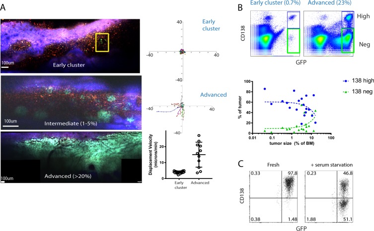
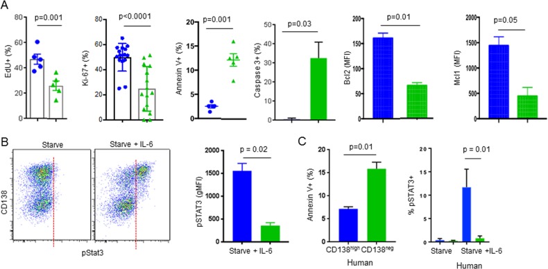
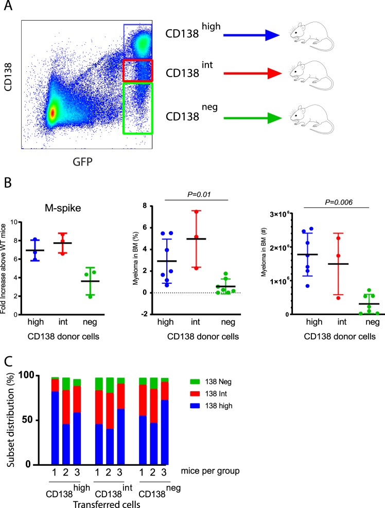
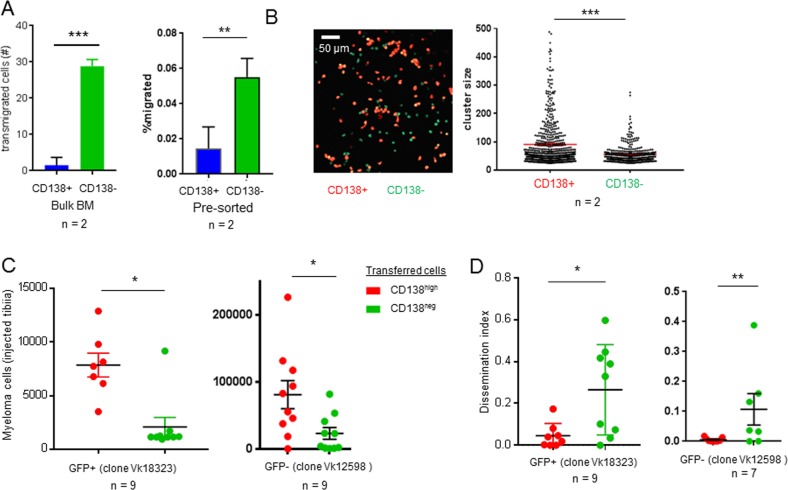
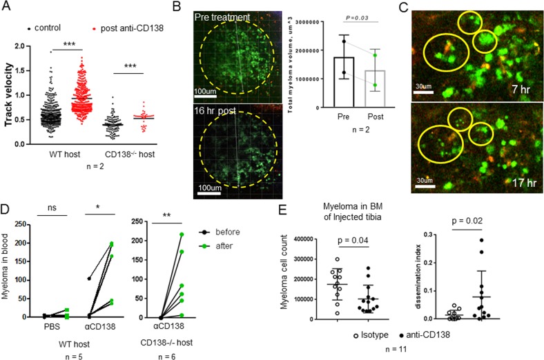
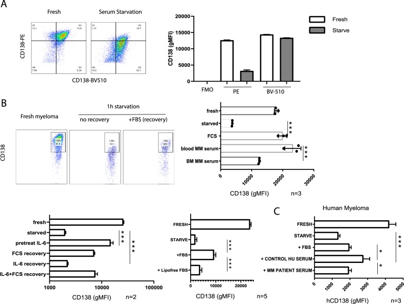
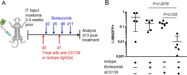
The canonical plasma cell marker CD138 (syndecan-1) is highly expressed on the myeloma cell surface, but its functional role in vivo is unclear, as well as the ontogeny of CD138-high and CD138-negative (neg) myeloma cells. In this study we used an in vivo murine Vk*MYC myeloma model where CD138 is heterogeneously expressed depending on tumor size. We find that in comparison to CD138-neg myeloma cells, the CD138-high subset of myeloma cells is highly proliferative, less apoptotic, and enhanced IL-6R signaling, which is known to promote survival. In addition CD138-high myeloma engrafts better than its CD138-neg counterpart. In contrast, CD138-neg cells are more motile both in vitro and in vivo, and more readily disseminate and spread to other bones in vivo than CD138-high subset. Neutralizing CD138 rapidly triggers migration of myeloma cells in vivo and leads to intravasation, which results in increased dissemination to other bones. Both murine and human myeloma cells can rapidly recycle CD138 surface expression through endocytic trafficking, in response to serum levels. Blocking CD138 enhances myeloma sensitivity to bortezomib chemotherapy and significantly reduces tumor size compared to bortezomib treatment alone. Thus, our data show that CD138 surface expression dynamically regulates a switch between growth vs. dissemination for myeloma, in response to nutrient conditions.


Conflict of interest statement

The authors declare that they have no conflict of interest.

Figures

Fig. 1
Fig. 1
CD138 expression and cell motility changes with tumor burden. a Intravital two-photon imaging of GFP+ (green) Vk*myc myeloma cells in the tibia at various tumor burdens (early, intermediate, and advanced stages) with analysis of individual cell track trajectories of total GFP+ myeloma cells in the BM, arrested at early stage cluster (yellow box in (a)) and representative migrating cells taken from advanced stage (>20% tumor burden) using mixed tumor of GFP+ and GFP-clones. b Representative dot plots of CD138 expression on myeloma cells (gated in black) at low and high tumor burden and overall trends in CD138-high and negative subsets plotted over various tumor sizes. c Comparison of CD138 expression on GFP+ myeloma cells either freshly isolated from BM, or after 1 h serum starvation (representative plot)
Fig. 2
Fig. 2
CD138 expression correlates with cell survival, proliferation, and IL-6 receptor signaling. a Comparison of CD138-high (blue), and negative (or neg in green) myeloma GFP+ subsets for proliferation, apoptosis, and survival factors from ex vivo fresh tumor, pre-gated on live cells. n is shown on each graph. b Freshly isolated myeloma was treated with or without exogenous IL-6 and IL-6 receptor signaling assessed by pSTAT3 intracellular staining on CD138 subsets for murine cells (n = 3). c Comparison of CD138-high and CD138-neg human myeloma (CD19− CD20− CD56+ CD138-high) cells by Annexin V and pSTAT3 (n = 3). All experiments were independently repeated two or more times. Mann–Whitney t tests were used. Errors reflect SD
Fig. 3
Fig. 3
CD138 expression promotes tumor engraftment and is dynamic in vivo. a Experimental design: FACS-purified populations of CD138 subsets (color coded) were i.v. transferred into sublethally irradiated (300Rad) WT recipients and analyzed 8 weeks later. b Total tumor burden was assessed by serum M-spike anti-IgG2b ELISA, and flow cytometry in the BM shown (frequency and in total cell counts). c Analysis of CD138 expression on myeloma from cohorts of mice show no difference in distribution or expression. Data are representative of two independent experiments (n = 3 mice per group per experiment). Mann–Whitney t tests were used. Errors reflect SD
Fig. 4
Fig. 4
CD138-neg myeloma cells are more motile in vitro and disseminate better in vivo. a Myeloma cell motility assessed by transwell assay. Percent frequency of CD138+ and CD138− myeloma cells in lower chamber using bulk tumor or FACS sorted and separately tested CD138+ and CD138− subsets (n = 2). b Sorted and labeled CD138+ (red) and CD138− (green) fractions were imaged on collagen coated glass, and cluster sizes were measured by image analysis software. c Analysis of tumor size in the injected (left) tibia 5 weeks after IT injection of FACS-purified CD138 subsets using GFP+ (Vk14451 clone) or GFP− (Vk12598 clone) from ex vivo serum-starved tumors. Analysis of dissemination index (ratio of tumor burden in contralateral tibia over injected tibia) using both clones. Data in de are pooled from two experiments for GFP+ clone and one GFP− experiment shown that is representative of the second one as well. Means were analyzed by Mann–Whitney t test. Errors reflect SEM in (a), and SD in (e)
Fig. 5
Fig. 5
Anti-CD138 rapidly mobilizes myeloma cells in the BM and promotes dissemination. a WT or CD138−/− recipient mice IT-injected myeloma, were imaged in the tibia, prior to (or untreated) with anti-CD138 (iv) or following treatment (2–6 h post) by time-lapse imaging. Myeloma cell motility was measured and pooled from two mice per group. b Representative images and comparisons of tumor cluster volume (by image analysis of GFP intensity) pre and 17 h post anti-CD138 treatment. c Images from time-lapse movies following treatment with circles highlighting apoptotic bodies. d Analysis of intravasation myeloma in blood prior to and following anti-CD138 treatment, PBS treatment in WT mice or in CD138−/− hosts. e Analysis of tumor burden in injected bone and dissemination index following chronic anti-CD138 or control antibody treatment for 4 weeks. n = 11. Errors reflect SD
Fig. 6
Fig. 6
CD138 surface expression is reversibly controlled by lipoproteins in serum. Freshly isolated myeloma cells were co-stained with PE anti-CD138 and BV510 anti-CD138, then serum starved 1 h and analyzed by flow cytometry. a Representative dot plots comparing PE and BV510 fluorescence before and after starvation, quantifying fluorescence intensity (geometric mean) for both fluorophores with FMO control, representative of two independent experiments. b Serum-starved myeloma recovers CD138 expression following re-culturing for 2 h with 10% FBS containing media, or other recovery conditions. c Human CD138 re-expression following starvation and serum recovery. Replicates pooled from independent experiments and analyzed by t tests. *p < 0.05, ***p < 0.001
Fig. 7
Fig. 7
Anti-CD138 enhances sensitivity to Bortezomib treatment. a Schematic of experimental design and treatment conditions. b Comparison of tumor burden in tibia, n = 5 mice per condition. Mann–Whitney t test analysis conducted. Errors reflect SD

References

    1. Kumar SK, Dispenzieri A, Lacy MQ, Gertz MA, Buadi FK, Pandey S, et al. Continued improvement in survival in multiple myeloma: changes in early mortality and outcomes in older patients. Leukemia. 2014;28:1122–8. doi: 10.1038/leu.2013.313. - DOI - PMC - PubMed
    1. Boise LH, Kaufman JL, Bahlis NJ, Lonial S, Lee KP. The Tao of myeloma. Blood. 2014;124:1873–9. doi: 10.1182/blood-2014-05-578732. - DOI - PMC - PubMed
    1. Fairfield H, Falank C, Avery L, Reagan MR. Multiple myeloma in the marrow: pathogenesis and treatments. Ann N Y Acad Sci. 2016;1364:32–51. doi: 10.1111/nyas.13038. - DOI - PMC - PubMed
    1. Di Marzo L, Desantis V, Solimando AG, Ruggieri S, Annese T, Nico B, et al. Microenvironment drug resistance in multiple myeloma: emerging new players. Oncotarget. 2016;7:60698–711. doi: 10.18632/oncotarget.10849. - DOI - PMC - PubMed
    1. Roccaro AM, Sacco A, Purschke WG, Moschetta M, Buchner K, Maasch C, et al. SDF-1 inhibition targets the bone marrow niche for cancer therapy. Cell Rep. 2014;9:118–28. doi: 10.1016/j.celrep.2014.08.042. - DOI - PMC - PubMed

Publication types