. 2019 Aug 7:13:181.
doi: 10.3389/fnbeh.2019.00181. eCollection 2019.

Sex-Dependent Modulation of Acute Stress Reactivity After Early Life Stress in Mice: Relevance of Mineralocorticoid Receptor Expression

Affiliations

Sex-Dependent Modulation of Acute Stress Reactivity After Early Life Stress in Mice: Relevance of Mineralocorticoid Receptor Expression

Valeria Bonapersona et al. Front Behav Neurosci. .

Abstract

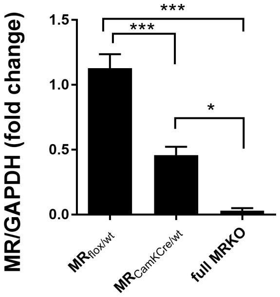
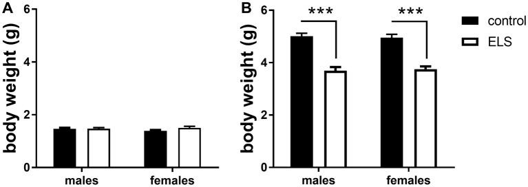
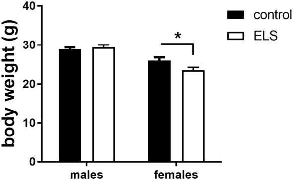
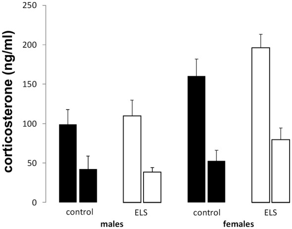
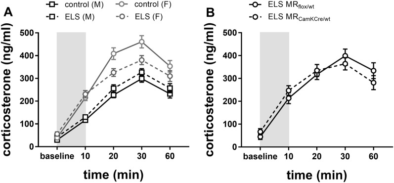
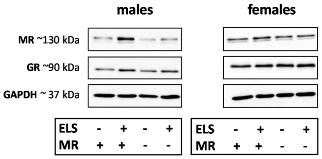
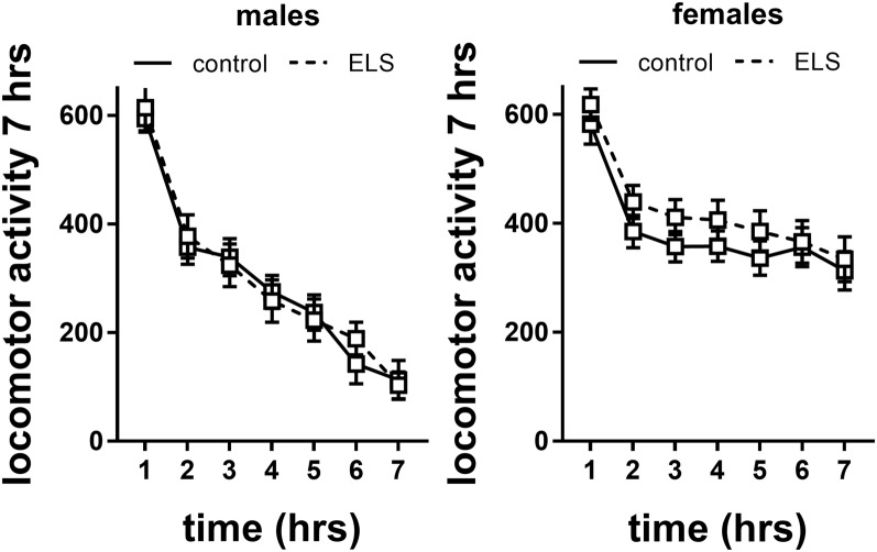
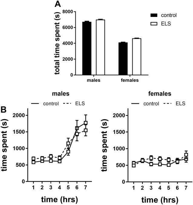
Early life stress (ELS) is considered a major risk factor for developing psychopathology. Increasing evidence points towards sex-dependent dysregulation of the hypothalamic-pituitary-adrenal (HPA) axis as a contributing mechanism. Additionally, clinical studies suggest that the mineralocorticoid receptor (MR) may further confer genetic vulnerability/resilience on a background of ELS. The link between ELS, sex and the HPA axis and how this interacts with MR genotype is understudied, yet important to understand vulnerability/resilience to stress. We used the early life-limited nesting and bedding model to test the effect of ELS on HPA properties in adult female and male mice carrying a forebrain-specific heterozygous knockout for MR. Basal HPA axis activity was measured by circadian peak and nadir corticosterone levels, in addition to body weight and weight of stress-sensitive tissues. HPA axis reactivity was assessed by mapping corticosterone levels after 10 min immobilization. Additionally, we measured the effects of ELS on steroid receptor [MR and glucocorticoid receptor (GR)] levels in the dorsal hippocampus and medial prefrontal cortex (mPFC) with western blot. Finally, behavioral reactivity towards a novel environment was measured as a proxy for anxiety-like behavior. Results show that HPA axis activity under rest conditions was not affected by ELS. HPA axis reactivity after immobilization was decreased by ELS in females and increased, at trend-level in males. This effect in females was further exacerbated by low expression of the MR. We also observed a sex*ELS interaction regarding MR and GR expression in the dorsal hippocampus, with a significant upregulation of MR in males only. The sex-dependent interaction with ELS was not reflected in the behavioral response to novel environment and time spent in a sheltered compartment. We did find increased locomotor activity in all groups after a history of ELS, which attenuated after 4 h in males but not females regardless of condition. Our findings support that ELS alters HPA axis functioning sex-dependently. Genetic predisposition to low MR function may render females more susceptible to the harmful effect of ELS whereas in males low MR function promotes resilience. We propose that this model may be a useful tool to investigate the underlying mechanisms of sex-dependent and genetic vulnerability/resilience to stress-related psychopathology.

Keywords: HPA axis; behavior; corticosterone; early life stress (ELS); mineralocorticoid receptor; neuroendocrine; nuclear receptors; sex.


Figures

Figure 1
Figure 1
Western blot analysis of mineralocorticoid receptor (MR) expression in the dorsal hippocampus of male MRflox/wt (n = 9), MRCamKCre/wt (n = 9) and full MRKO mice (n = 3). Significant differences across groups, after Holm’s post hoc analysis, are indicated: ***p < 0.001, *p < 0.05. Data presented as mean ± SEM.
Figure 2
Figure 2
Averaged body weight (±SEM) of the pups in experimental litters (A) at the start (P2; B) and end (P9) for control (black bars) and early life stress (ELS) condition (open bars). Mean weight per litter was calculated for both males and females. Data were analyzed with averaged litter size at P2 as covariate for the analysis of body weight at P9. Significance indicates main effect of ELS (F(1,44) = 89.45, p < 0 0.001), ***p < 0.001.
Figure 3
Figure 3
Averaged body weight (±SEM) of adult control (black bars) and ELS animals (open bars). Mean weight per group (±SEM) was calculated for both males and females. Significant differences between groups using a t-test with Holm’s correction are indicated, *p < 0.05.
Figure 4
Figure 4
Peak (left bar) and nadir (right bar) plasma corticosterone levels in all experimental groups. Plasma corticosterone was unaffected by ELS (open bars) vs. control (black bars) in both males and females; n = 22–24 per group. Data presented as mean ± SEM.
Figure 5
Figure 5
(A) Plasma corticosterone levels after immobilization (10 min restraint stress) were analyzed using repeated measures over time in control males and females (black and gray solid line) or in ELS males and females (black and gray dotted line; n = 22–24 per group). Data presented as mean ± SEM. (B) Plasma corticosterone levels after immobilization plotted for genotype in the females exposed to ELS (n = 9–11 per group). Data presented as mean ± SEM.
Figure 6
Figure 6
Representative images of MR, glucocorticoid receptor (GR) and GAPDH western blots in male and female samples of the dorsal hippocampus (n = 5–9 per experimental group). The results of the medial prefrontal cortex (mPFC) were highly comparable. Legends in the graph: ELS (− or +) refers to either control or ELS condition; MR (− or +) refers to the MRflox/wt or MRCamKCre/wt.
Figure 7
Figure 7
Locomotor activity in males and females exposed to control or ELS condition. Data analyzed with repeated measures ANOVA. n = 28–34 per group in males; n = 22–26 per group in females. Locomotor activity = linear combination of standardized distance moved, velocity and time spent moving. Data presented as mean ± SEM.
Figure 8
Figure 8
Time spent in the shelter of the home cages in the first 7 h of exposure to a novel environment expressed as (A) total time spent and (B) over time for males and females exposed to either control or ELS condition (n = 18–22 per group in males; n = 18–23 per group in females. Data presented as mean ± SEM.

Similar articles

Cited by

References

    1. Andersen S. L. (2003). Trajectories of brain development: point of vulnerability or window of opportunity? Neurosci. Biobehav. Rev. 27, 3–18. 10.1016/s0149-7634(03)00005-8 - DOI - PubMed
    1. Arp J. M., ter Horst J. P., Kanatsou S., Fernández G., Joëls M., Krugers H. J., et al. . (2014). Mineralocorticoid receptors guide spatial and stimulus-response learning in mice. PLoS One 9:e86236. 10.1371/journal.pone.0086236 - DOI - PMC - PubMed
    1. Bale T. L. (2006). Stress sensitivity and the development of affective disorders. Horm. Behav. 50, 529–533. 10.1016/j.yhbeh.2006.06.033 - DOI - PubMed
    1. Bale T. L., Epperson C. N. (2015). Sex differences and stress across the lifespan. Nat. Neurosci. 18, 1413–1420. 10.1038/nn.4112 - DOI - PMC - PubMed
    1. Barbazanges A., Piazza P. V., Le Moal M., Maccari S. (1996). Maternal glucocorticoid secretion mediates long-term effects of prenatal stress. J. Neurosci. 16, 3943–3949. 10.1523/jneurosci.16-12-03943.1996 - DOI - PMC - PubMed

LinkOut - more resources