. 2019 Aug 1;10(4):756-769.
doi: 10.14336/AD.2018.0912. eCollection 2019 Aug.

Glial S100A6 Degrades β-amyloid Aggregation through Targeting Competition with Zinc Ions

Affiliations

Glial S100A6 Degrades β-amyloid Aggregation through Targeting Competition with Zinc Ions

Zhi-Ying Tian et al. Aging Dis. .

Abstract

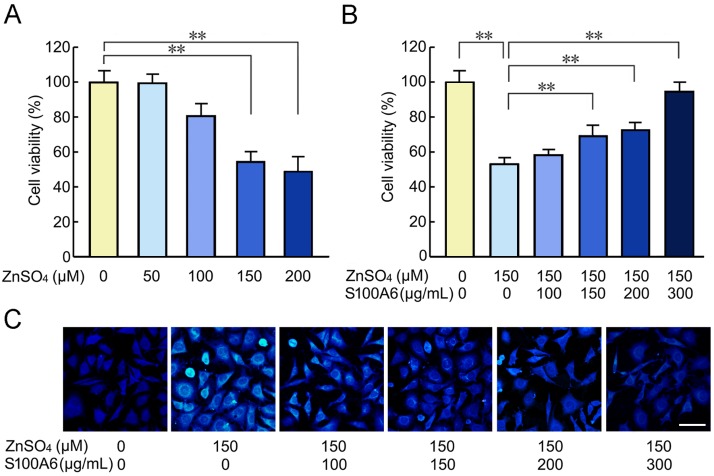
Evidence has been accumulating that zinc ions can trigger β-amyloid (Aβ) deposition and senile plaque formation in the brain, a pathological hallmark of Alzheimer's disease (AD). Chelating zinc inhibits Aβ aggregation and may hold promise as a therapeutic strategy for AD. S100A6 is an acidic Ca2+/Zn2+-binding protein found only in a small number of astrocytes in the normal brain. However, in the AD brain, S100A6 is highly expressed in astrocytes around Aβ plaques. The role of the astrocytic S100A6 upregulation in AD is unknown. In the present study, we examined the effects of S100A6 on Aβ plaques and intracellular zinc levels in a mouse model of AD. Chronic exposure to zinc increased Aβ deposition and S100A6 expression, both reversible by the zinc chelator clioquinol, in the brains of amyloid precursor protein/presenilin 1 (APP/PS1) transgenic mice. To examine whether exogenous S100A6 could induce Aβ plaque disaggregation through competition for zinc in vitro, we incubated APP/PS1 mouse brain sections with recombinant human S100A6 protein or co-incubated them with human S100A6-expressing cells. Both treatments efficiently reduced the Aβ plaque burden in situ. In addition, treatment with exogenous S100A6 protected cultured COS-7 cells against zinc toxicity. Our results show for the first time that increased S100A6 levels correlate with both Aβ disaggregation and decrease of Aβ plaque-associated zinc contents in brain sections with AD-like pathology. Astrocytic S100A6 in AD may protect from Aβ deposition through zinc sequestration.

Keywords: Alzheimer’s disease; S100A6; astrocyte; zinc; β-amyloid protein.


Conflict of interest statement

Conflict of Interest We have no conflicting interest to disclose.

Figures

Figure 1.
Figure 1.. Confocal images showing the effects of zinc and chelator treatments on S100A6 expression and Aβ aggregation in the APP/PS1 mouse brain
The 9-month-old APP/PS1 mice on high-zinc (Zn), zinc + clioquinol (Zn + CQ), or CQ diet were sacrificed. Age-matched APP/PS1 mice given a standard diet and deionized water were used as the controls (Con). (A) Frozen sections of the brain double immunostained with anti-Aβ (green) and anti-S100A6 (red) antibodies showing the distribution and expression of Aβ (a1-d1) and S100A6 (a2-d2), and their co-localization (a3-d3). Aβ and S100A6 immunostaining show significant co-localization in the brain sections of APP/PS1 mice on a high-zinc diet. The Y-Z images depicted in the right panel indicate the positive immunofluorescence staining after orthogonal sectioning. (B) The intensity of Aβ plaques and Aβ plaques-associated S100A6 immunofluorescence were determined. (C) Atomic absorption spectrum assay was used for the measurement of zinc levels in the cortex. Values are means ± S.E.M. Results were compared by a two-way ANOVA followed by t test (n = 5). **P < 0.01. Scale bars = 20 μm.
Figure 2.
Figure 2.. High-zinc diet led to an increase of S100A6 protein expression in the brains of APP/PS1 mice
RT-PCR (A, B) and Western blot (C, D) assay were used to detect the mRNA and protein levels of S100A6, respectively, in the brains of APP/PS1 mice fed with high-zinc, zinc + CQ, or CQ diet. Age-matched APP/PS1 mice administered with standard diet and deionized water served as controls (Con). The results are presented as percentages, and the control is defined as 100%. Values represent means ± S.E.M. Results were compared by a two-way ANOVA followed by t test, **P < 0.01 versus the controls, ## P < 0.01 versus the zinc treatment group (n = 5).
Figure 3.
Figure 3.. Recombinant human S100A6 (hS100A6) protein reversed zinc-induced cell toxicity
The MTT assay results show the changes in the cell viability with the addition of indicated concentrations of zinc sulfate (ZnSO4) (A) and different concentrations of hS100A6 protein along with 150 μM of ZnSO4 (B). Cells treated with the vehicle served as controls. Zinquin staining was used to detect zinc in the cells that were incubated with 150 μM ZnSO4 and different concentrations of hS100A6 protein (C). Values are means ± S.E.M. and are representative of at least three independent experiments. Results were compared by one-way ANOVA with post-hoc Fisher’s protected least significant difference (PLSD) test, **P < 0.01. Scale bars = 30 μm.
Figure 4.
Figure 4.. Overexpression of hS100A6 mitigated zinc-induced decrease of COS-7 cell viability
COS-7 cells were transfected with pcDNA3.1-hS100A6 before being incubated with zinc sulfate (0 µM, 50 µM, 100 µM, 150 µM and 200 µM) for 12 h. COS-7 cells transfected with pcDNA3.1 vector were used as controls (Con). (A) The S100A6 mRNA levels were evaluated using RT-PCR at 48 and 72 h after transfection. (B) Western blot analyses demonstrated S100A6 protein overexpression in the transfected COS-7 cells and in controls. The levels of S100A6 mRNA and protein were expressed as the ratio of the mean intensity at indicated time to the level at 24 h after transfection. (C) Immunofluorescence staining for S100A6 indicate the representative images S100A6-positive cells (Arrows) confirmed the successful transfection of COS-7 with pcDNA3.1-hS100A6 (c1: 0 h; c2: 24 h; c3: 48h; c4: 72h). (D) MTT and lactate dehydrogenase (LDH) assays were performed to determine cell viability after addition of the indicated concentrations of zinc sulfate. The control was defined as 100%. (E) Intracellular zinc levels were detected using the Zinquin staining. Values are means ± S.E.M. and are representative of at least three independent experiments. Results were compared by one-way ANOVA with post-hoc Fisher’s protected least significant difference (PLSD) test, *P < 0.05; **P < 0.01. Scale bars = 30 μm.
Figure 5.
Figure 5.. Recombinant hS100A6 protein contributed to Aβ degradation in brain slices of the APP/PS1 mouse
Brain slices collected from APP/PS1 mouse were incubated in Dulbecco’s Modified Eagle Medium (DMEM) supplemented with recombinant hS100A6 protein for 24 h. Adjacent sections from the same brains incubated in DMEM medium alone were used as controls (Con). Immunohistochemical staining with anti-Aβ antibody demonstrated Aβ protein expression. Representative images indicating the Aβ deposits in the cerebral cortex (A) and hippocampus (B). High-magnification images of representative Aβ-positive staining are shown in the right panels. (C) The Aβ plaque burden were quantified. (D) Aβ42 levels were determined by ELISA assay. The content of Aβ42 was expressed as ng per mg of tissue protein. (E) Thioflavin-S (Thio S) and N-(6-methoxy-8-quinolyl)-p-toluenesulfonamide (TSQ) stainings showed the distribution of fibrillar Aβ and zinc, respectively, in the slices of APP/PS1 mouse brain incubated with hS100A6 protein or medium alone. Values are means ± S.E.M. Results were compared by Student’s t test (n = 4). *P < 0.05, **P < 0.01 versus the controls. Scale bars: A, B = 200 μm, and 20 μm in the high magnification of right panels; D = 20 μm.
Figure 6.
Figure 6.. Overexpression of hS100A6 was associated with the clearance of amyloid plaques in brain sections of the APP/PS1 mice
Brain slices and adjacent sections from the same brains collected from APP/PS1 mouse were co-incubated with COS-7 cells transfected with pcDNA3.1-hS100A6 or with pcDNA3.1-hS100A6 empty vector (control, Con), respectively, for 24 h. Representative immunohistochemistry images showing the Aβ plaques in the cerebral cortex (A) and hippocampus (B) of the APP/PS1 mice brain. The right panels depict representative Aβ plaques in high magnification. Quantification of Aβ burden demonstrated a significant decrease in the sections incubated with COS-7 cells expressing hS100A6 compared to controls (C). (D) TSQ fluorescence staining was performed to detect zinc levels in the slices of APP/PS1 mouse brain co-incubated with cells expressing hS100A6 or controls. Values are means ± S.E.M. Results were compared by Student’s t test (n = 4). *P < 0.05, **P < 0.01 versus the controls. Scale bars: A, B = 200 μm, and 20 μm in the high magnification right panels; D = 20 μm.

Similar articles

Cited by

References

    1. Eckerstrom C, Klasson N, Olsson E, Selnes P, Rolstad S, Wallin A (2018). Similar pattern of atrophy in early- and late-onset Alzheimer's disease. Alzheimers Dement (Amst), 10: 253-259. - PMC - PubMed
    1. Ding Q, Tanigawa K, Kaneko J, Totsuka M, Katakura Y, Imabayashi E, et al. (2018). Anserine/Carnosine Supplementation Preserves Blood Flow in the Prefrontal Brain of Elderly People Carrying APOE e4. Aging Dis, 9: 334-345. - PMC - PubMed
    1. Guglielmotto M, Monteleone D, Piras A, Valsecchi V, Tropiano M, Ariano S, et al. (2014). Abeta1-42 monomers or oligomers have different effects on autophagy and apoptosis. Autophagy, 10: 1827-1843. - PMC - PubMed
    1. Szybinska A, Lesniak W (2017). P53 Dysfunction in Neurodegenerative Diseases - The Cause or Effect of Pathological Changes? Aging Dis, 8: 506-518. - PMC - PubMed
    1. Bearer EL, Manifold-Wheeler BC, Medina CS, Gonzales AG, Chaves FL, Jacobs RE (2018). Alterations of functional circuitry in aging brain and the impact of mutated APP expression. Neurobiol Aging, 70: 276-290. - PMC - PubMed

LinkOut - more resources