. 2019 Aug 7:9:286.
doi: 10.3389/fcimb.2019.00286. eCollection 2019.

Influenza Virus Infection Induces ZBP1 Expression and Necroptosis in Mouse Lungs

Affiliations

Influenza Virus Infection Induces ZBP1 Expression and Necroptosis in Mouse Lungs

Yun Wang et al. Front Cell Infect Microbiol. .

Abstract

Programmed cell death and especially necroptosis, a programmed and regulated form of necrosis, have been recently implicated in the progression and outcomes of influenza in mouse models. Moreover, Z-DNA/RNA binding protein 1 (ZBP1) has been identified as a key signaling molecule for necroptosis induced by Influenza A virus (IAV). Direct evidence of IAV-induced necroptosis has not been shown in infected lungs in vivo. It is also unclear as to what cell types undergo necroptosis during pulmonary IAV infection and whether ZBP1 expression can be regulated by inflammatory mediators. In this study, we found that IAV infection induced ZBP1 expression in mouse lungs. We identified that mediators implicated in the pathogenesis of IAV infection including interferons (IFNs), TNFα, and agonists for Toll-like receptors 3 and 4 were potent inducers of ZBP1 expression in primary murine alveolar epithelial cells, bone marrow derived macrophages, and dendritic cells. We further found that IAV infection induced a strong necroptosis through phosphorylation of the necroptosis effector mixed lineage kinase domain-like protein in infiltrating immune cells and alveolar epithelial cells by day 7 post-infection. Lastly, we found different cell type-specific responses to IAV-induced cell death upon inhibition of caspases and/or necroptosis pathways. Our findings provide direct evidence that IAV infection induces necroptosis in mouse lungs, which may involve local induction of ZBP1 and different programmed cell death signaling mechanisms in alveolar epithelial and infiltrating inflammatory cells in the lungs.

Keywords: Influenza A virus; ZBP1; lung; necroptosis; programmed cell death.


Figures

Figure 1
Figure 1
Western blot detection of ZBP1 and necroptosis in IAV-infected mouse lungs. C57BL/6J mice were infected intranasally with a lethal dose (1,000 pfu/mouse, ~2 LD50) of the mouse-adapted IAV H1N1 PR/8/34 strain or treated with saline as a negative control (Con). (A) Mouse lungs were harvested on days 6 and 7 post-infection and lung homogenates at equal protein amounts were subjected to Western blot analysis with indicated antibodies. (B) Bronchoalveolar lavage cells from control and IAV-infected mice on day 3 (d3) post-infection were collected and equal amounts of cell lysates were subjected to Western blot analysis with indicated antibodies. Results represent the findings of three independent experiments.
Figure 2
Figure 2
Immunohistochemical detection of ZBP1 in IAV-infected mouse lungs. Lung sections from saline-treated (Mock, A,B) or H1N1 PR8-infected mice on day 7 post-infection (C,D) were subjected to immunostaining with a ZBP1 monoclonal antibody by using Vector M.O.M immunodetection kits (n = 5). Positive immunoreactivities (red) were detected in the alveolar epithelial cells (red arrows) and immune cells (green arrows). The region indicated in (C) (magnification, x100) is shown at higher magnification in (D) (x400).
Figure 3
Figure 3
Induction of ZBP1 by IAV infection and immune stimuli in primary mouse alveolar epithelial cells and immune cells. (A,B) Primary mAECs were treated with control PBS (- or control), infected with active H1N1 PR/8/34 strain at MOI of 5, 10, or 20, or treated with mouse TNFα (20 ng/ml), IFNα2 (50 ng/ml), IFNγ (50 ng/ml), TNFα plus IFNα2 (T+α2, 20 + 50 ng/ml), TNFα plus IFNγ (T+γ, 20 + 50 ng/ml), GM-CSF (30 ng/ml), GM-CSF plus IFNα2 (GM+α2, 30 + 50 ng/ml), GM-CSF plus IFNγ (GM+γ, 30 + 50 ng/ml) for 24 h. (C–F) BMDMs, BMDCs and RAW264.7 murine macrophages were treated with control PBS (mock or control), infected with H1N1 PR/8/34, H3N2 A/Hong Kong/8/68 (H3N2-HK), or H3N2 (x:31) A/Aichi/68 strains at MOI of 5, or treated with mouse IFNα2 (50 ng/ml), IFNγ (50 ng/ml), TNFα (20 ng/ml), LPS (100 ng/ml), poly(I:C) (PIC, 1 μg/ml), TNFα plus IFNα2 (T+α2, 20 + 50 ng/ml), or TNFα plus IFNγ (T+γ, 20 + 50 ng/ml) for 24 h. Equal amounts of cell lysates from (A–F) were subjected to Western blotting with indicated antibodies. Results represent the findings of three independent experiments. The production of IFNβ by 24 h treatment of LPS (100 ng/ml) and poly(I:C) (PIC, 1 μg/ml) was shown in the bar graph of (D) (n = 3).
Figure 4
Figure 4
IAV induces phosphorylation of MLKL in alveolar epithelial cells and infiltrated immune cells in infected mouse lungs. Lung sections from saline-treated (Mock, A,B) or PR8-infected mice on day 7 post-infection (C–F) were subjected to immunostaining with a phospho-MLKL (Ser345) monoclonal antibody using a Vector M.O.M immunodetection kit (n = 6). Positive p-MLKL immunoreactivities (red) were detected in the alveolar epithelial cells (red arrows) and immune cells (green arrows). The region indicated in (E) (magnification, x100) is shown at higher magnification in (F) (x400). Final magnification for (A) is x100 and for (B–D) x400. In (A–D), the lung sections were from inflated mouse lungs by intratracheal infusion of 10% formalin to total lung capacity.
Figure 5
Figure 5
Immunofluorescence detection of IAV-induced phosphorylation of MLKL and its co-localization with pulmonary infiltrated immune cells. (A,B) Lung sections from control (A) or PR8-infected mice (B) on day 7 post-infection were subjected to immunofluorescence staining with a phospho-MLKL (Ser345) monoclonal antibody by using Vector M.O.M immunodetection kit followed by Alexa Fluor 568-labeled goat anti-mouse secondary IgG (n = 6). Positive p-MLKL immunoreactivities are stained red (indicated by green arrows) and cell nuclei stained blue by DAPI. Final magnification is x400. (C,D) Lung sections from PR8-infected mice on day 7 post-infection were subjected to double immunofluorescence staining with the phospho-MLKL (Ser345) mouse monoclonal antibody, Alexa Fluor-488 conjugated CD45 or FITC conjugated CD192/CCR2 rat monoclonal antibodies as described in section Materials and Methods. Positive p-MLKL immunoreactivities are stained red, and CD45 or CCR2 are stained green. The co-localization or overlay is shown as yellow and indicated by white arrows. Final magnification: x400 (n = 6).
Figure 6
Figure 6
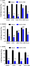
Cell type-specific responses to IAV-induced cell death upon inhibition of caspases and/or RIPK3. Primary mAECs, BMDMs, and BMDCs were treated with control PBS (Mock) or infected with H1N1 PR8 at 5 MOI in the presence of DMSO, v-ZVD-FMK (ZVD, 60 μM), GSK872 (GSK, 5 μM), v-ZVD-FMK plus GSK872 (ZVD+GSK), or RIPK1 inhibitor II (Nec-1s, 5 μM) for 24 h (B,C) or 48 h (A). Cell viability was assessed by MTS assay (CellTiter Aqueous One Solution Assay) and cell survival rates were calculated by comparison to DMSO-treated control mock cells and are presented as means ± SE (n = 3). NS, no significance; *p < 0.05; **p < 0.01; ***p < 0.001 vs. DMSO. Cell survival rates relative to individual mock control groups were also shown over the bar as indicated.

References

    1. Atkin-Smith G. K., Duan M., Chen W., Poon I. K. H. (2018). The induction and consequences of Influenza A virus-induced cell death. Cell Death Dis. 9:1002. 10.1038/s41419-018-1035-6 - DOI - PMC - PubMed
    1. Cho Y. S., Challa S., Moquin D., Genga R., Ray T. D., Guildford M., et al. . (2009). Phosphorylation-driven assembly of the RIP1-RIP3 complex regulates programmed necrosis and virus-induced inflammation. Cell 137, 1112–1123. 10.1016/j.cell.2009.05.037 - DOI - PMC - PubMed
    1. Downey J., Pernet E., Coulombe F., Allard B., Meunier I., Jaworska J., et al. . (2017). RIPK3 interacts with MAVS to regulate type I IFN-mediated immunity to Influenza A virus infection. PLoS Pathog. 13:e1006326. 10.1371/journal.ppat.1006326 - DOI - PMC - PubMed
    1. Downey J., Pernet E., Coulombe F., Divangahi M. (2018). Dissecting host cell death programs in the pathogenesis of influenza. Microbes Infect. 20, 560–569. 10.1016/j.micinf.2018.03.005 - DOI - PMC - PubMed
    1. Estabragh Z. R., Mamas M. A. (2013). The cardiovascular manifestations of influenza: a systematic review. Int. J. Cardiol. 167, 2397–2403. 10.1016/j.ijcard.2013.01.274 - DOI - PubMed

Publication types

MeSH terms