. 2019 Jul 29:11:100218.
doi: 10.1016/j.bonr.2019.100218. eCollection 2019 Dec.

Stat3 in osteocytes mediates osteogenic response to loading

Affiliations

Stat3 in osteocytes mediates osteogenic response to loading

Kylie A Corry et al. Bone Rep. .

Abstract

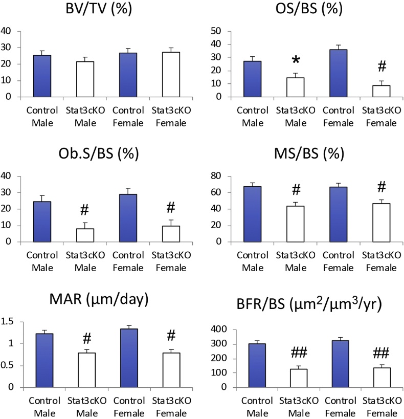
Signal transducer and activator of transcription 3 (Stat3) is a member of the Stat family of proteins involved in signaling in many different cell types, including osteocytes. Osteocytes are considered major mechanosensing cells in bone due to their intricate dendritic networks able to sense changes in physical force and to orchestrate the response of osteoclasts and osteoblasts. We examined the role of Stat3 in osteocytes by generating mice lacking Stat3 in these cells using the Dmp-1(8kb)-Cre promoter (Stat3cKO mice). Compared to age-matched littermate controls, Stat3cKO mice of either sex (18 weeks old) exhibit reduced bone formation indices, decreased osteoblasts and increased osteoclasts, and altered material properties, without detectable changes in bone mineral density (BMD) or content of either trabecular or cortical bone. In addition, Stat3cKO mice of either sex show significantly decreased load-induced bone formation. Furthermore, pharmacologic inhibition of Stat3 in osteocytes in vitro with WP1066 blocked the increase in cytosolic calcium induced by ATP, a mediator of the cellular responses to sheer stress. WP1066 also increased reactive oxygen species (ROS) production in cultured MLO-Y4 osteocytes. These data demonstrate that Stat3 is a critical mediator of mechanical signals received by osteocytes and suggest that osteocytic Stat3 is a potential therapeutic target to stimulate bone anabolism.

Keywords: ATP; Bone formation; Mechanotransduction; Osteocyte; ROS; Signal transducers and activators of transcription 3 (Stat3).


Conflict of interest statement

The authors declare that there is no conflict of interest.

Figures

Fig. 1
Fig. 1
Osteocytes specific Stat3 knockout leads to decreased Stat3 protein expression in osteocytes and lower bone mass compared to WT mice. Immunohistochemical staining of experimental mice cortical bone of femurs. Immunohistochemical staining demonstrates the expression of Stat3 protein in 8-week-old osteocytes of the Dmp1-Cre; Stat3+/+ (WT control) mouse (A1). Stat3 proteins in osteocytes were stained brown and dark brown. However, there was no Stat3 protein expression in osteocytes of the Dmp1-Cre; Stat3flox/flox (conditional Stat3 KO) mouse (B1). Black arrows indicate osteocytes without Stat3 proteins probed by brown stains in conditional Stat3 KO mouse (Scale bar = 25 μm). Three-dimensional images of distal femur trabecular bones show, compared to Dmp1-Cre;Stat3+/+ (WT) mouse trabecular bones (A2), the Dmp1-Cre;Stat3flox/flox (KO) mouse trabecular bones (B2) demonstrated no significant difference in trabecular bone number and volume. Trabecular bone in the distal femur with fluorescent labels (calcein and alizarin) in (A3) Dmp1-Cre;Stat3+/+ (WT) and (B3) Dmp1-Cre;Stat3flox/flox (KO) mice, indicating less labels in conditional Stat3 KO mice.
Fig. 2
Fig. 2
Stat3 cKO mice exhibit lower BV/TV and decreased bone formation. Histomorphometric measurements of trabecular bone in the distal femur indicate bone volume (BV/TV), Osteoid surface (OS/BS), Osteoblast surface (Ob.S./BS), mineralizing surface (MS/BS), mineral appositional rate (MAR) and bone formation rate (BFR/BS). Note that *: p < 0.05, #: p < 0.01 and ##: p < 0.001 versus the same sex control.
Fig. 3
Fig. 3
Stat3 cKO mice exhibit higher bone resorption than the WT control. TRAP stained osteoclasts on trabecular bone surfaces in distal femurs. Compared to Dmp1-Cre;Stat3+/+ (WT) mouse (A), Dmp1-Cre;Stat3flox/flox (KO) mouse (B) presented more osteoclast number and osteoclast surface (red stain on trabecular bone surface) on trabecular bone in the similar region of distal femur 0.5 mm above the growth plate. Scale bar = 50 μm. Osteoclast surface (Oc.S/BS) of osteocyte-specific Stat3 KO mice were 177% greater in males and 110% greater in females compared to their controls. Osteoclast number (N.Oc/B.Pm) of osteocyte-specific Stat3 KO mice were 146% greater in males and 104% greater in females compared to their controls.
Fig. 4
Fig. 4
Stat3 deficiency in osteocytes negatively affects biomechanical properties of cortical bone. The three point bending test of femoral shafts shows that ultimate force, stiffness, and energy to failure were not significantly different between the wild type controls and Dmp1-Cre;Stat3flox/flox (KO) mice. Material properties based on the geometric data measured showed that ultimate stress and Young's modulus were significantly less in Dmp1-Cre;Stat3flox/flox (KO) mice, compared to the wild type controls. Toughness was not different between the wild type controls and Dmp1-Cre;Stat3flox/flox (KO) mice. Note that #: p < 0.01 and ##: p < 0.001.
Fig. 5
Fig. 5
Stat3 deficiency in osteocytes suppresses mechanically induced bone formation. Midshaft ulnar sections were acquired from the control and loaded forearms for the wild type controls and Dmp1-Cre;Stat3flox/flox (KO) mice. Calcein (green) and alizarin (red) injections were given after loading. Note that the anabolic responses on the medial (square) and lateral surfaces of the loaded control ulna were detected, but those responses were significantly decreased in the loaded ulna of the Dmp1-Cre;Stat3flox/flox (KO) mice. Bone morphometric parameters for the control mice and Dmp1-Cre;Stat3flox/flox (KO) mice include relative mineralizing surface (rMS/BS), relative mineral appositional rate (rMAR) and relative bone formation rate (rBFR/BS). Note that ##: p < 0.001.
Fig. 6
Fig. 6
The effect of external ATP on cytosolic Ca2+ concentration ([Ca2+]c) in cultured osteocytes (A, C-F) and osteoblasts (B). In A and B, concentration dependence of ATP-induced Ca2+ transients in osteocytes and osteoblasts, respectively. Numbers in parentheses show CaCl2 concentrations in μM. Here, horizontal lines indicate the applied treatment (i.e. ATP application). In C, Ca2+ transients in osteocytes triggered by repeated application of external ATP (10 μM, 30 s). In D, 1 μM FCCP inhibited ATP-induced Ca2+ transients in osteocytes triggered by 10 μM ATP. Then, ATP was removed by washing cells with ATP-free solution. Fig. 7C shows the dependence of ATP effects on external Ca2+. Where indicated, osteocytes were treated with 10 μM ATP and exposed to 1.8 mM CaCl2 or Ca2+-free solution as indicated. In D, thapsigargin (0.1 μM) inhibited Ca2+ transients in osteocytes triggered by 10 μM ATP.
Fig. 7
Fig. 7
Inhibition of Stat3 function suppressed cytosolic Ca2+ concentration ([Ca2+]c) caused by external ATP and increased ROS production in cultured osteocytes. In A, similar to Fig. 7C, Ca2+ transients in osteocytes triggered by repeated application of external ATP (10 μM, 30 s). WP1066, an inhibitor of Stat3, suppressed Ca2+ transients triggered by external ATP in osteocytes (B). In B, where indicated, 10 μM ATP, 10 or 50 μM WP1066, were applied to osteocytes. Numbers in parentheses indicate concentrations of the inhibitor in μM. In C, ROS production in vehicle-treated osteocytes. In D, WP1066 increased ROS production in osteocytes. Cells were loaded with dihydroethidium (DHE, 5 μM) and then pre-incubated with a vehicle (0.05% DMSO, E) or 10 μM WP1066 (F) for 30 min. The vehicle (0.05% DMSO) and WP1066 (10 μM) remained in the culture solution during the experiment.

References

    1. Aaronson D.S., Horvath C.M. A road map for those who don⬢t know JAK-STAT. Science. 2002;296(5573):1653–1655. - PubMed
    1. Almeida M., Han L., Martin-Millan M., O⬢Brien C.A., Manolagas S.C. Oxidative stress antagonizes Wnt signaling in osteoblast precursors by diverting beta-catenin from T cell factor- to forkhead box O-mediated transcription. J. Biol. Chem. 2007;282(37):27298–27305. - PubMed
    1. Bakker A.D., Kulkarni R.N., Klein-Nulend J., Lems W.F. 2014. IL-6 Alters Osteocyte Signaling Toward Osteoblasts but Not Osteoclasts; pp. 394–399. - PubMed
    1. Berman A.G., Wallace J.M., Bart Z.R., Allen M.R. Raloxifene reduces skeletal fractures in an animal model of osteogenesis imperfecta. Matrix Biol. 2016;52–54:19–28. - PubMed
    1. Bivi N., Condon K.W., Allen M.R., Farlow N., Passeri G., Brun L.R., Rhee Y., Bellido T., Plotkin L.I. Cell autonomous requirement of connexin 43 for osteocyte survival: consequences for endocortical resorption and periosteal bone formation. Journal of bone and mineral research: the official journal of the American Society for Bone and Mineral Research. 2012;27(2):374–389. - PMC - PubMed

LinkOut - more resources