

Fibroblasts: Diverse Cells Critical to Biomaterials Integration
- PMID: 31440581
- PMCID: PMC6705602
- DOI: 10.1021/acsbiomaterials.7b00244
Fibroblasts: Diverse Cells Critical to Biomaterials Integration
Abstract
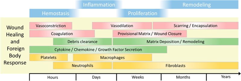
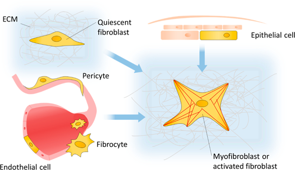
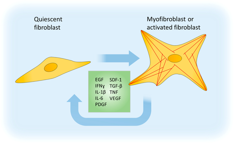
Fibroblasts are key participants in wound healing and inflammation, and are capable of driving the progression of tissue repair to fully functional tissue or pathologic scar, or fibrosis, depending on the specific mechanical and biochemical cues with which they are presented. Thus, understanding and modulating the fibroblastic response to implanted materials is paramount to achieving desirable outcomes, such as long-term implant function or tissue regeneration. However, fibroblasts are remarkably heterogeneous and can differ vastly in their contributions to regeneration and fibrosis. This heterogeneity exists between tissues and within tissues, down to the level of individual cells. This review will discuss the role of fibroblasts, the pitfalls of describing them as a collective, the specifics of their function, and potential future directions to better understand and organize their highly variable biology.
Keywords: biomaterials; fibroblast; fibrosis; inflammation; mechanobiology; mechanotransduction; review; wound healing.
Figures
References
-
- Atluri P The Surgical Review: An Integrated Basic and Clinical Science Study Guide; Lippincott Williams & Wilkins, 2005; p 300.
-
- Robbins S; Cotran RS Acute and Chronic Inflammation In Pathologic Basis of Disease; Saunders: Philadelphia, PA, 1989; pp 47–86.
Grants and funding
LinkOut - more resources
Full Text Sources