

Giant renal vein aneurysm
- PMID: 31440715
- PMCID: PMC6699197
- DOI: 10.1016/j.jvscit.2019.06.007
Giant renal vein aneurysm
Abstract
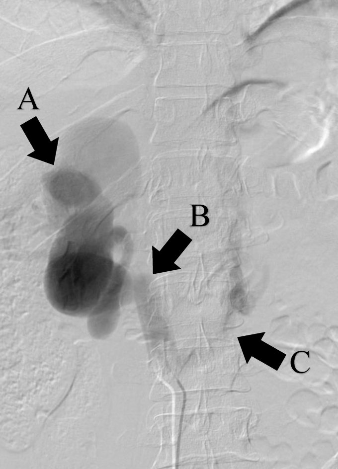
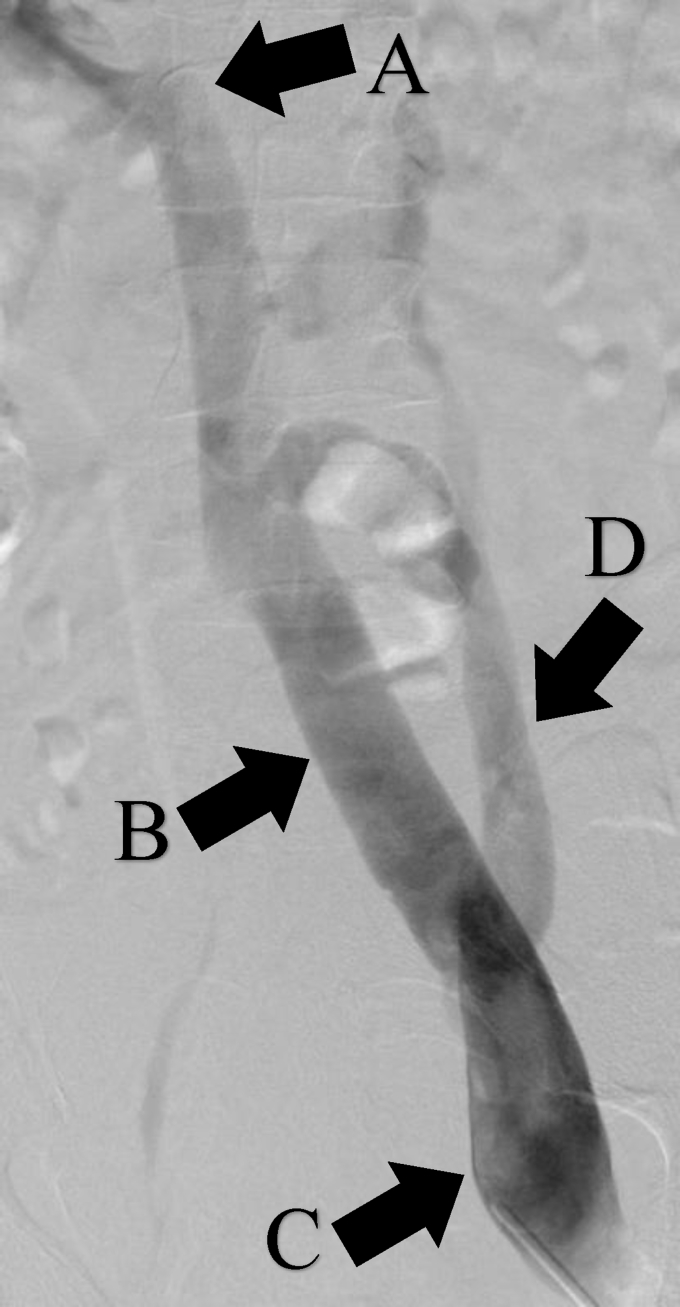
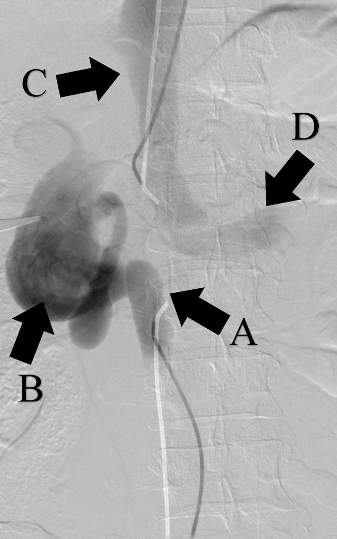
A 37-year-old asymptomatic man presented with incidentally identified intra-abdominal venous aneurysms. Workup, which included venography, demonstrated an absent segment of the inferior vena cava between the inferior right and superior left renal vein, resulting in a 4.4-cm right renal vein aneurysm, dilated common iliac veins, and left external iliac vein aneurysm. Collateralization was robust. Given the limited natural history data and complexities of open reconstruction, we opted to observe this asymptomatic patient with serial imaging, which demonstrated no interval change. We present our case and a review of the literature pertaining to intra-abdominal venous aneurysms.
Keywords: Absent IVC; Renal vein aneurysm; Venous aneurysms.
Figures


References
-
- Teter K.A., Maldonado T.M., Adelman M.A. A systematic review of venous aneurysms by anatomic location. J Vasc Surg Venous Lymphat Disord. 2018;6:408–413. - PubMed
-
- Laurenzi A., Ettorre G.M., Lionetti R., Meniconi R.L., Colasanti M., Vennarecci G. Portal vein aneurysm: what to know. Dig Liver Dis. 2015;47:918–923. - PubMed
-
- Calligaro K.D., Ahmad S., Dandora R., Dougherty M.J., Savarese K.J., Doerr K.J. Venous aneurysms: surgical indications and review of the literature. Surgery. 1995;117:1–6. - PubMed
-
- Park J.S., Kim J.Y., Kim M., Park S.C., Lee K.Y., Won Y.S. Ruptured aneurysm of the external iliac vein. J Vasc Surg Venous Lymphat Disord. 2016;4:92–94. - PubMed
-
- Gradman W.S., Steinberg F. Aneurysm of the inferior vena cava: case report and review of the literature. Ann Vasc Surg. 1993;7:347–353. - PubMed
Publication types
LinkOut - more resources
Full Text Sources
