. 2020 May;14(3):373-384.
doi: 10.1007/s12072-019-09977-w. Epub 2019 Aug 22.

Wnt status-dependent oncogenic role of BCL9 and BCL9L in hepatocellular carcinoma

Affiliations

Wnt status-dependent oncogenic role of BCL9 and BCL9L in hepatocellular carcinoma

Nicole Huge et al. Hepatol Int. 2020 May.

Abstract

Background: Activation of Wnt/β-catenin pathway is a frequent event in hepatocellular carcinoma and is associated with enhanced cell survival and proliferation. Therefore, targeting this signaling pathway is discussed as an attractive therapeutic approach for HCC treatment. BCL9 and BCL9L, two homologous coactivators of the β-catenin transcription factor complex, have not yet been comprehensively characterized in HCC. We aimed to elucidate the roles of BCL9 and BCL9L, especially regarding Wnt/β-catenin signaling and their prognostic value in HCC.

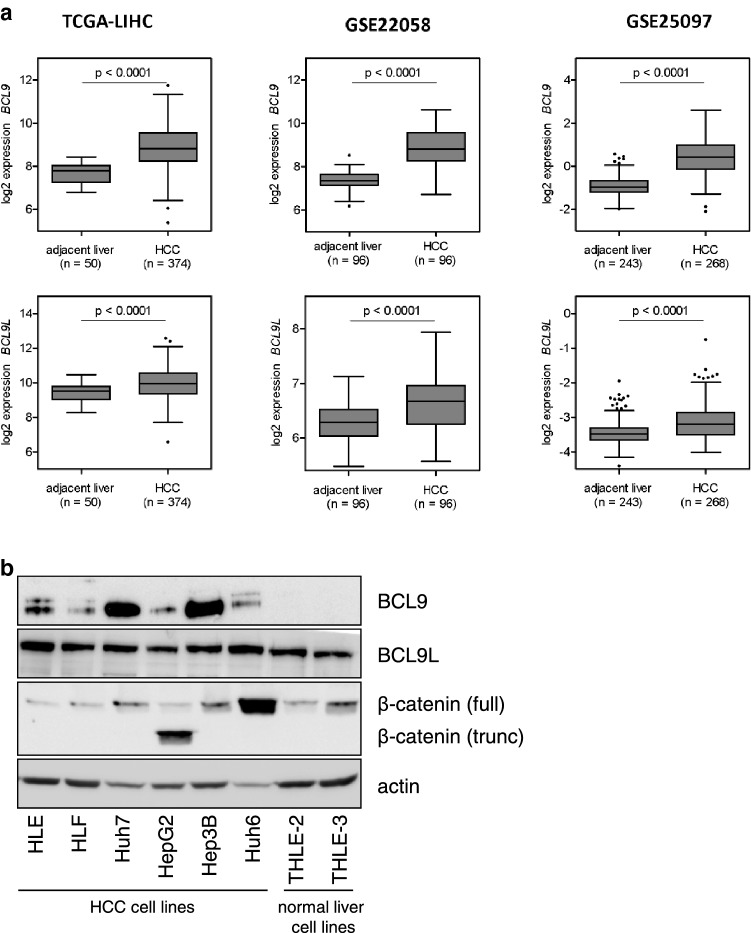
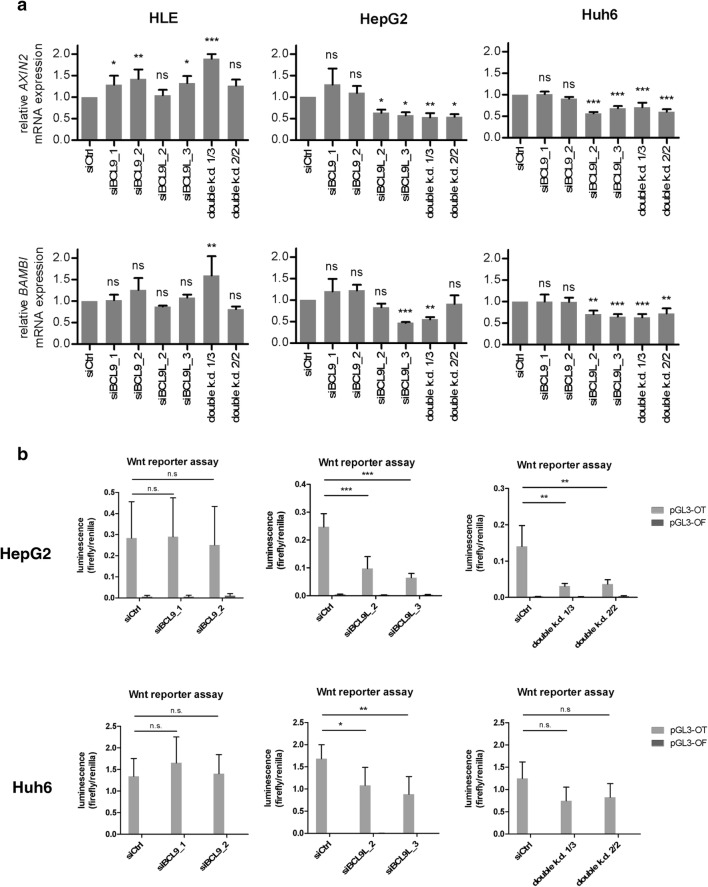
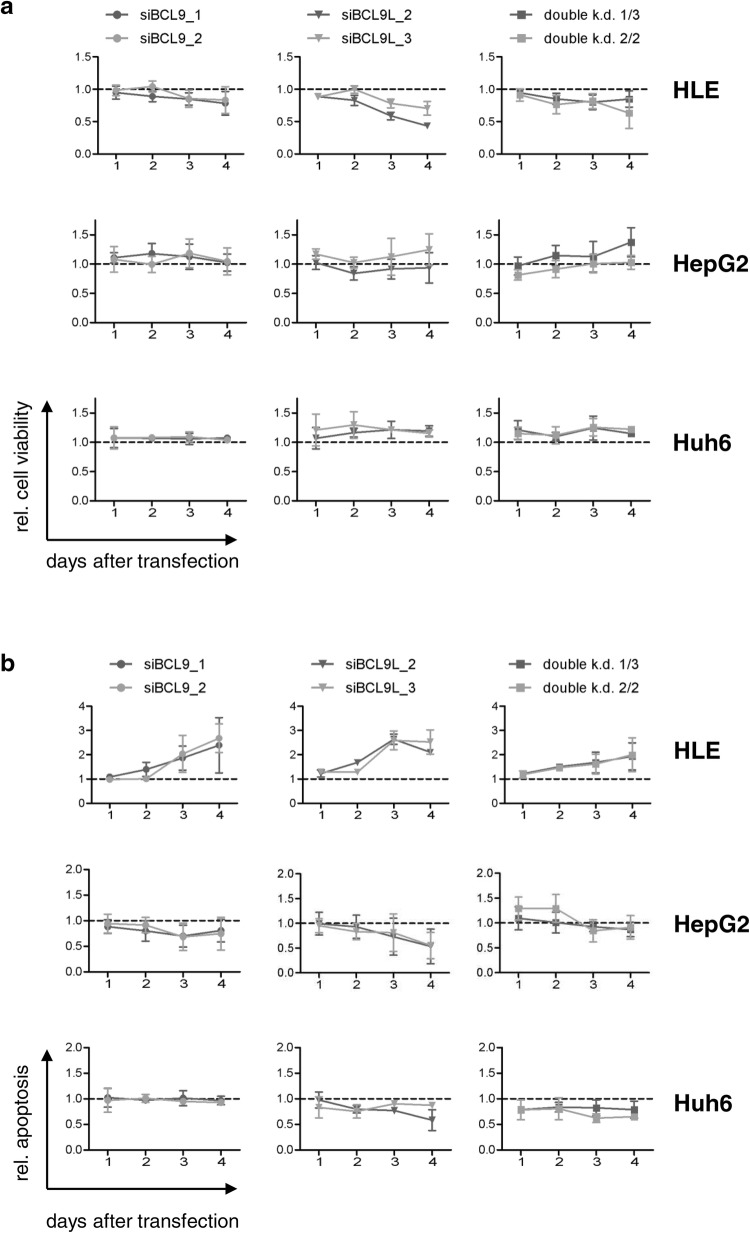
Methods: Expression of BCL9/BCL9L was determined in HCC cell lines (HLE, HLF, Huh7, HepG2, Hep3B, and Huh6) and normal liver cell lines (THLE-2 and THLE-3). To analyze proliferation and apoptosis, BCL9 and/or BCL9L were knocked down in Wnt-inactive HLE and Wnt-active HepG2 and Huh6 cells using siRNA. Subsequently, Wnt reporter assays were performed in HepG2 and Huh6 cells. BCL9 and BCL9L expression, clinicopathological and survival data of public HCC datasets were analyzed, taking the Wnt signaling status into account.

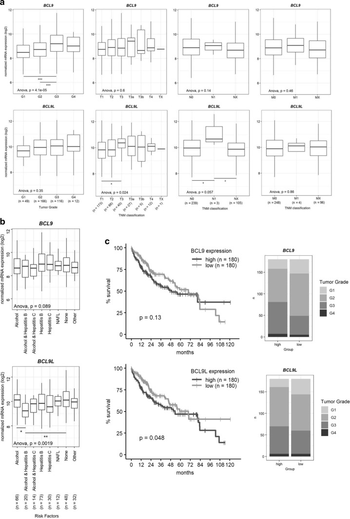
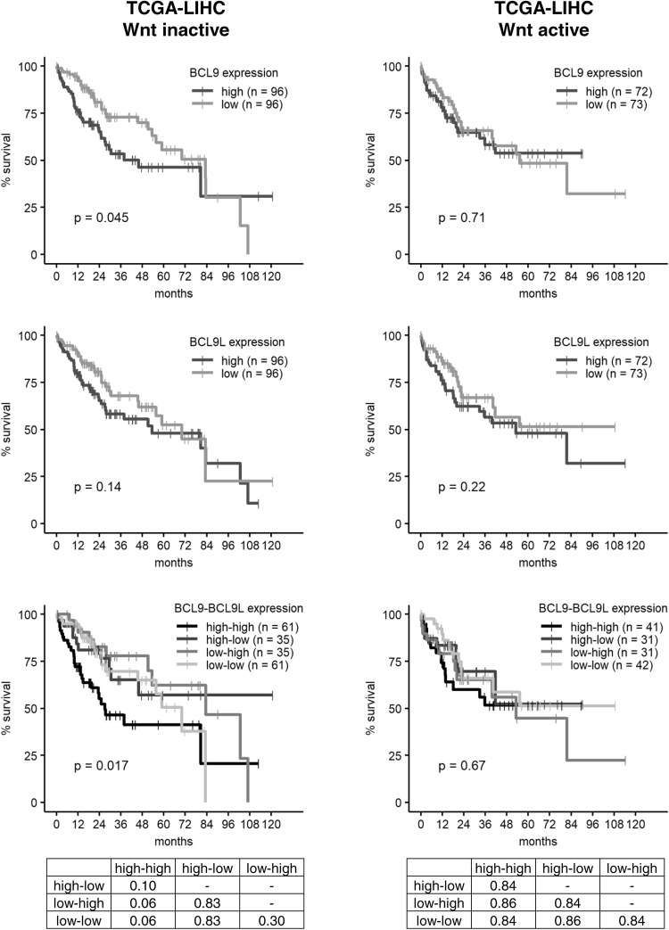
Results: Knockdown of BCL9L, but not of BCL9, reduced Wnt signaling activity. Knockdown of BCL9 and/or BCL9L reduced cell viability and increased apoptosis of Wnt-inactive HCC cells, but had no effect in Wnt-active cells. Expression of BCL9 and BCL9L was upregulated in human HCC and increased with progressing dedifferentiation. For BCL9L, higher expression was observed in tumors of larger size. Overexpression of BCL9 and BCL9L correlated with poor overall survival, especially in HCC without activated Wnt signaling.

Conclusion: Oncogenic BCL9 proteins represent promising targets for cancer therapy and inhibiting them may be particularly beneficial in Wnt-inactive HCCs.

Keywords: B9L; BCL9-2; Liver cancer.


Conflict of interest statement

The authors declare that they have no conflict of interest.

Figures

Fig. 1
Fig. 1
BCL9 and BCL9L are overexpressed in human HCC. aBCL9 and BCL9L expression levels were analyzed using the three public HCC data sets TCGA-LIHC, GSE22058, and GSE25097. BCL9 and BCL9L expression was significantly higher in HCC tissue than in adjacent non-tumorous liver tissue. Tukey box-and-whisker plot; two-tailed Student’s t test. b Western blot analysis of HCC cell lines (HLE, HLF, Huh7, HepG2, Hep3B, and Huh6) and immortalized liver cell lines (THLE-2, THLE-3) for BCL9, BCL9L, and β-catenin protein expression. HepG2 and Huh6 harbor an activating β-catenin deletion and mutation, respectively, causing an accumulation of β-catenin
Fig. 2
Fig. 2
BCL9 and BCL9L overexpression correlates with poor survival of HCC patients. aBCL9 and BCL9L expression levels of the TCGA-LIHC cohort were divided according to histologic tumor grade and TNM classification parameters [27]. Data are represented as Tukey’s box-and-whisker plot. *p < 0.05, ***p < 0.001; 1-way ANOVA with Tukey’s multiple comparison test. bBCL9 and BCL9L expression levels of the TCGA-LIHC cohort were divided according to tumor etiology. Data are represented as Tukey’s box-and-whisker plot. *p < 0.05, **p < 0.01; one-way ANOVA with Tukey’s multiple comparison test. cBCL9 and BCL9L expression values and survival data of the TCGA-LIHC cohort were retrieved from http://www.oncolnc.org/ [16]. Patients were grouped into low (lower median) or high (upper median) expressions of BCL9 or BCL9L. Kaplan–Meier with log-rank test
Fig. 3
Fig. 3
Knockdown of BCL9L or BCL9/BCL9L interrupts Wnt/β-catenin signaling, while knockdown of BCL9 alone is insufficient to decrease Wnt/β-catenin activity. a Expression analysis of AXIN2 and BAMBI by qRT-PCR using the ΔΔCT method in HLE, HepG2, and Huh6 cells after transfection with siBCL9 and/or siBCL9L. Data are represented as mean ± SD of three independent experiments. *p < 0.05, **p < 0.01, ***p < 0.001, n.s. not significant; one-way ANOVA with Dunnett’s multiple comparison test. b Effects of BCL9 and/or BCL9L knockdown on Wnt/β-catenin signaling activity of HepG2 and Huh6 cells was determined by Wnt reporter assays. Cells were co-transfected with combinations of siBCL9 and/or siBCL9L and pGL3-OT or pGL3-OF. For normalization, the renilla luciferase vector pGL4.70 was used in all conditions. Data are represented as mean ± SD of at least three independent experiments. *p < 0.05, **p < 0.01, ***p < 0.001, ns not significant; one-way ANOVA with Tukey’s multiple comparison test
Fig. 4
Fig. 4
Knockdown of BCL9 and/or BCL9L prevents proliferation and induces apoptosis in HLE cells, but not in HepG2 or Huh6 cells. HLE, HepG2, and Huh6 cells were transfected with 5 nM siRNA against BCL9 and/or BCL9L. Cell viability and apoptosis were determined every 24 h. Data are represented as mean ± SD of at least three independent experiments. a Cell viability was analyzed by WST-1 assay and normalized to siControl (dotted line). b Apoptosis was analyzed by caspase 3/7 activity and normalized to cell viability and siControl (dotted line)
Fig. 5
Fig. 5
High levels of BCL9 and BCL9L correlate with poor survival only in HCCs with inactive Wnt signaling. BCL9 and BCL9L expression values and survival data of the TCGA-LIHC cohort were retrieved from http://www.oncolnc.org/ [16] and stratified into Wnt-inactive and Wnt-active HCCs according to Sanchez-Vega et al. [17]. BCL9 and BCL9L expression was classified as high (upper median) or low (lower median) and, for combined survival analysis, patients were grouped into the following BCL9BCL9L expression groups: high–high, high–low, low–high, and low–low. The tables show the corresponding p values for the combined BCL9BCL9L survival analysis as determined by log-rank test with Benjamini–Hochberg correction for multiple testing. Survival was analyzed according to Kaplan–Meier

Similar articles

Cited by

References

    1. Stewart BW, Wild CP. World Cancer Report 2014. Lyon: International Agency for Research on Cancer/World Health Organization. 2014
    1. Llovet JM, Ricci S, Mazzaferro V, Hilgard P, Gane E, Blanc J-F, et al. Sorafenib in advanced hepatocellular carcinoma. N Engl J Med. 2008;359:378–390. doi: 10.1056/NEJMoa0708857. - DOI - PubMed
    1. Kudo M, Finn RS, Qin S, Han K-H, Ikeda K, Piscaglia F, et al. Lenvatinib versus sorafenib in first-line treatment of patients with unresectable hepatocellular carcinoma: a randomised phase 3 non-inferiority trial. Lancet. 2018;391:1163–1173. doi: 10.1016/S0140-6736(18)30207-1. - DOI - PubMed
    1. Schulze K, Imbeaud S, Letouzé E, Alexandrov LB, Calderaro J, Rebouissou S, et al. Exome sequencing of hepatocellular carcinomas identifies new mutational signatures and potential therapeutic targets. Nat Genet. 2015;47:505–511. doi: 10.1038/ng.3252. - DOI - PMC - PubMed
    1. Khalaf AM, Fuentes D, Morshid AI, Burke MR, Kaseb AO, Hassan M, et al. Role of Wnt/β-catenin signaling in hepatocellular carcinoma, pathogenesis, and clinical significance. J Hepatocell Carcinoma. 2018;5:61–73. doi: 10.2147/JHC.S156701. - DOI - PMC - PubMed

MeSH terms