. 2019 Oct;7(10):e00860.
doi: 10.1002/mgg3.860. Epub 2019 Aug 23.

MiR-497-5p inhibits cell proliferation and metastasis in hepatocellular carcinoma by targeting insulin-like growth factor 1

Affiliations

MiR-497-5p inhibits cell proliferation and metastasis in hepatocellular carcinoma by targeting insulin-like growth factor 1

Guo-Shu Xu et al. Mol Genet Genomic Med. 2019 Oct.

Retraction in

Abstract

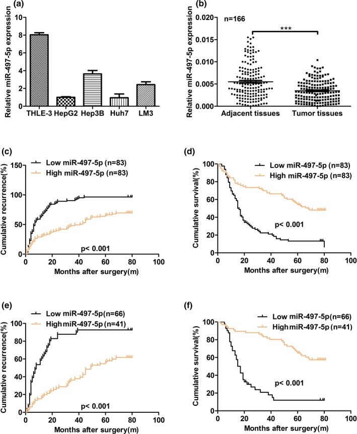
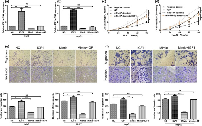
Background: MicroRNAs (miRNAs) play an important regulatory role in carcinogenesis and cancer progression. Aberrant expression of miR-497-5p has been reported in various human malignancies. However, the role of miR-497-5p in hepatocellular carcinoma (HCC) remains unclear.

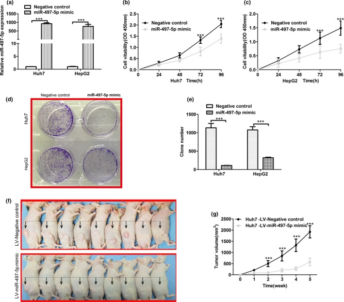
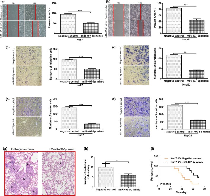
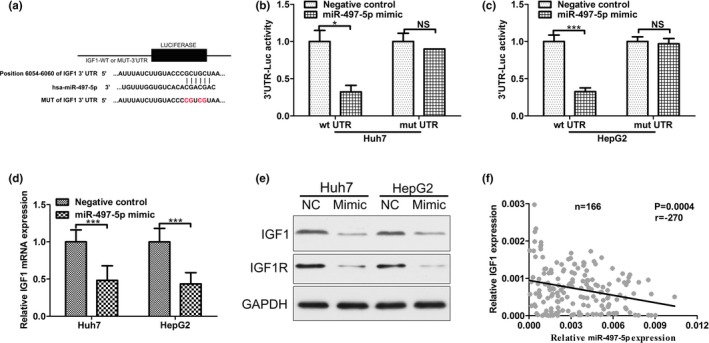
Results: In this study, we found that miR-497-5p was downregulated in HCC tissues. The low level of miR-497-5p in HCC tumors was correlated with aggressive clinicopathological characteristics and predicted poor prognosis in HCC patients. The overexpression of miR-497-5p significantly inhibited HCC cell proliferation, colony formation, and metastasis in vitro and vivo. Bioinformatics analysis further identified insulin-like growth factor 1 (IGF1) as a novel target of miR-497-5p in HCC cells.

Conclusion: Our study suggested that miR-497-5p regulates HCC cell survival, partially through downregulation of IGF1. Therefore, the miR-497-5p/IGF1 axis might serve as a novel therapeutic target in patients with HCC.

Keywords: IGF1; biomarker; hepatocellular carcinoma; miR-497-5p; proliferation.


Conflict of interest statement

We certify that the authors have no actual or potential conflict of interest in relation to this article.

Figures

Figure 1
Figure 1
miR‐497‐5p expression is downregulated in human hepatocellular carcinoma (HCC). (a) The expression of miR‐497‐5p was significantly lower in HCC cell lines compared with normal liver cells. miR‐497‐5p expression was normalized to U6 expression. (b) The relative mRNA expression of miR‐497‐5p was analyzed in 166 cases of HCC using real‐time polymerase chain reaction and normalized to U6 expression. (c) The low miR‐497‐5p expression group showed a shorter recurrence free survival than the high miR‐497‐5p expression group. (d) The low miR‐497‐5p expression group showed a shorter overall survival than the high miR‐497‐5p expression group. (e, f) The prognostic value of miR‐497‐5p was also observed in patients with early‐stage HCC (TNM stage I). Statistical significance was assessed by two‐sided log‐rank tests. ***p < .001
Figure 2
Figure 2
Overexpression of miR‐497‐5p inhibits hepatocellular carcinoma cell growth in vitro and in vivo. (a) Stable overexpression of miR‐497‐5p in Huh7 and HepG2 cells, respectively, were established using a lentiviral system and assessed using quantitative real‐time polymerase chain reaction. MiR‐497‐5p expression was normalized to U6 expression. (b, c) MTT assays were performed 24, 48, 72, and 96 hr after transfection to determine the proliferation of Huh7 and HepG2 cells. Data represent the mean ± SD from three independent experiments. (d, e) Colony formation assays were performed in Huh7 and HepG2 cells transfected with miR‐497‐5p mimics or negative control miR‐GFP. The average number of colonies and representative images are shown. (f) Representative images and (g) tumor growth in xenografted mice 5 weeks after subcutaneous injection with either miR‐497‐5p or miR‐GFP cells (n = 9). ***p < .001
Figure 3
Figure 3
Overexpression of miR‐497‐5p inhibits hepatocellular carcinoma cell metastasis in vitro and vivo. (a, b) The migratory properties of miR‐497‐5p‐overexpressing cells and control cells were analyzed by scratch wound healing assays in Huh7 and HepG2 cells. Representative results are shown. Magnification: ×100. (c, d) The migratory properties of the cells were analyzed using the Transwell migration assay with Transwell filter chambers. Results are plotted as the average number of migrated cells from six random microscopic fields. (e, f) The invasive properties of the cells were analyzed with the invasion assay using BioCoat Matrigel invasion chambers. Results are plotted as the average number of invasive cells from six random microscopic fields. (g) Representative H&E images of mouse lung tissue sections from the LV‐miR‐497‐5p mimic and LV‐Negative control groups (magnification: ×200) black arrows indicated Lung neoplastic foci. (h) The number of metastatic foci in the lungs of each group (n = 9) is presented as the mean ± SD (error bars). (i) Comparisons of the overall survival curves of mice injected with either LV‐miR‐497‐5p mimic or LV‐Negative control. p‐values were calculated using the two‐sided log‐rank test. *p < .05; ***p < .001
Figure 4
Figure 4
MiR‐497‐5p directly targets insulin‐like growth factor 1 (IGF1). (a) The putative miR‐497‐5p binding sites in the 3’UTR of IGF1 mRNA are shown. A mutation was generated on the IGF1 3’UTR sequence in the complementary site for the seed sequence of miR‐497‐5p. (b) Wild‐type (IGF1 3’UTR‐WT) or mutant (IGF 3’UTR‐mut) reporter plasmids were co‐transfected into Huh7 cells with miR‐497‐5p or miR‐GFP. The normalized luciferase activity in the control group was set as the relative luciferase activity. (c) Wild‐type (IGF1 3’UTR‐WT) or mutant (IGF 3’UTR‐mut) reporter plasmids were co‐transfected into HepG2 cells with miR‐497‐5p or miR‐GFP. The normalized luciferase activity in the control group was set as the relative luciferase activity. (d) The mRNA expression of IGF1 was analyzed using RT‐PCR in Huh7 and HepG2. GAPDH was used as an internal control. (e) The protein expression of IGF1 and IGF1R was analyzed using western blotting in Huh7 and HepG2. GAPDH was used as an internal control. All experiments were performed in triplicate with similar results. (f) The correlation between the miR‐497‐5p level and IGF1 mRNA level was measured in the same set of tissues. *p < .05; ***p < .001
Figure 5
Figure 5
Rescue expression of insulin‐like growth factor 1 (IGF1) abolished the effects of miR‐497‐5p on phenotypes of HCC cells. (a, b) The expression of IGF1 in Huh7 and HepG2 cells, respectively, were examined by quantitative real‐time polymerase chain reaction. (c, d) MTT assays were performed 24, 48, 72, and 96 hr after transfection to determine the proliferation of Huh7 and HepG2 cells. Data represent the mean ± SD from three independent experiments. (e, f) The migratory properties of the cells were analyzed using the Transwell migration assay with Transwell filter chambers. Results are plotted as the average number of migrated cells from six random microscopic fields and the invasive properties of the cells were analyzed with the invasion assay using BioCoat Matrigel invasion chambers. Results are plotted as the average number of invasive cells from six random microscopic fields. *p < .05; **p < .01; ***p < .001

References

    1. Abba, M. , Mudduluru, G. , & Allgayer, H. (2012). MicroRNAs in cancer: Small molecules, big chances. Anti‐Cancer Agents in Medicinal Chemistry. 12(7):733‐743 - PubMed
    1. Bosch, F. X. , Ribes, J. , Díaz, M. , & Cléries, R. (2004). Primary liver cancer: Worldwide incidence and trends. Gastroenterology, 127(5), S5–S16. 10.1053/j.gastro.2004.09.011 - DOI - PubMed
    1. Brouwer‐Visser, J. , & Huang, G. S. (2015). IGF2 signaling and regulation in cancer. Cytokine & Growth Factor Reviews, 26(3), 371–377. 10.1016/j.cytogfr.2015.01.002 - DOI - PubMed
    1. Bruchim, I. , Attias, Z. , & Werner, H. (2009). Targeting the IGF1 axis in cancer proliferation. Expert Opinion on Therapeutic Targets, 13(10), 1179 10.1517/14728220903201702 - DOI - PubMed
    1. Chai, L. , Kang, X. J. , Sun, Z. Z. , Zeng, M. F. , Yu, S. R. , Ding, Y. , … Zhao, J. MiR‐497‐5p, miR‐195‐5p and miR‐455‐3p function as tumor suppressors by targeting hTERT in melanoma A375 cells. Cancer Management and Research. 2018;10:989‐1003. 10.2147/CMAR.S163335 - DOI - PMC - PubMed

Publication types

MeSH terms