. 2019 Aug 22;12(17):2669.
doi: 10.3390/ma12172669.

Sodium Alginate/Gelatine Hydrogels for Direct Bioprinting-The Effect of Composition Selection and Applied Solvents on the Bioink Properties

Affiliations

Sodium Alginate/Gelatine Hydrogels for Direct Bioprinting-The Effect of Composition Selection and Applied Solvents on the Bioink Properties

Dorota Bociaga et al. Materials (Basel). .

Abstract

Hydrogels tested and evaluated in this study were developed for the possibility of their use as the bioinks for 3D direct bioprinting. Procedures for preparation and sterilization of hydrogels and the speed of the bioprinting were developed. Sodium alginate gelatine hydrogels were characterized in terms of printability, mechanical, and biological properties (viability, proliferation ability, biocompatibility). A hydrogel with the best properties was selected to carry out direct bioprinting tests in order to determine the parameters of the bioink, adapted to print with use of the designed and constructed bioprinter and provide the best conditions for cell growth. The obtained results showed the ability to control mechanical properties, biological response, and degradation rate of hydrogels through the use of various solvents. The use of a dedicated culture medium as a solvent for the preparation of a bioink, containing the predicted cell line, increases the proliferation of these cells. Modification of the percentage of individual components of the hydrogel gives the possibility of a controlled degradation process, which, in the case of printing of temporary medical devices, is a very important parameter for the hydrogels' usage possibility-both in terms of tissue engineering and printing of tissue elements replacement, implants, and organs.

Keywords: bioink for scaffolds; bioprinting; cell viability; degradation of hydrogels; mechanical properties; micro-extrusion.


Conflict of interest statement

The authors declare no conflict of interest. The funders had no role in the design of the study; in the collection, analyses, or interpretation of data; in the writing of the manuscript, or in the decision to publish the results.

Figures

Figure 1
Figure 1
Rheological properties of the samples dissolved in water (W) (a) and full culture medium (M) (b) with different contents of gelatine.
Figure 2
Figure 2
Compressive strength results for the samples dissolved in water (W) and full culture medium (M) with different content of gelatine (* = p < 0.05 and ** = p < 0.01, results statistically significant).
Figure 3
Figure 3
Degradation rates for samples dissolved in water (W) and full culture medium (M) with different content of gelatine evaluated after 24 h, 48 h, 7 days, and 14 days of incubation (* = p < 0.05, results statistically significant).
Figure 4
Figure 4
Compressive strength results for samples dissolved in water (W) and full culture medium (M) with different content of gelatine evaluated after 24 h, 48 h, 7 days, and 14 days of incubation (* = p < 0.05, results statistically significant).
Figure 5
Figure 5
Compressive strength results for samples dissolved in water (W) and full culture medium (M) with different content of gelatine after 0 (0 C, reference samples), 10 (10 C), 20 (20 C), and 50 (50 C) cycles of loading and unloading (* = p < 0.05, results statistically significant).
Figure 6
Figure 6
Swelling ratio for samples dissolved in water (W) and full culture medium (M) with different content of gelatine measured after 1 h, 3 h, 6 h, 24 h, and 27 h.
Figure 7
Figure 7
Endothelial cells (EA.hy 926 line) viability after direct contact with samples dissolved in water (W) and full culture medium (M) with different content of gelatine after 24 and 48 h of incubation.
Figure 8
Figure 8
Endothelial cells (EA.hy 926 line) cytotoxicity after direct contact with 5A4GW and 5A4GM samples after 24 (a,c) and 48 h (b,d) of incubation (live cells are marked green, dead in red). Magnification 100x.
Figure 9
Figure 9
Endothelial cells (EA.hy 926 line) viability after indirect contact with samples dissolved in water (W) and full culture medium (M) with different content of gelatine after 24 and 48 h of incubation.
Figure 10
Figure 10
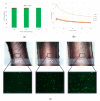
Influence of different temperatures of extrusion (34, 37, and 40 °C) on the endothelial cells (EA.hy 926 line) viability (a), viscosity (b), and path width (c) of the 5A4GM hydrogel. Photomicroscopy, 100× magnification, fluorescence microscopy, 50× magnification.

References

    1. Ullah F., Bisyrul M., Othman H., Javed F., Ahmad Z., Akil M. Classification, processing and application of hydrogels: A review. Mater. Sci. Eng. C. 2015;57:414–433. doi: 10.1016/j.msec.2015.07.053. - DOI - PubMed
    1. Akhtar M.F., Hanif M., Ranjha N.M. Methods of synthesis of hydrogel A review. Saudi Pharm. J. 2016;24:554–559. doi: 10.1016/j.jsps.2015.03.022. - DOI - PMC - PubMed
    1. Hoffman A.S. Hydrogels for biomedical applications. Adv. Drug Deliv. Rev. 2012;64:18–23. doi: 10.1016/j.addr.2012.09.010. - DOI - PubMed
    1. Chai Q., Jiao Y., Yu X. Hydrogels for Biomedical Applications: Their Characteristics and the Mechanisms behind Them. Gels (Basel Switz.) 2017;3:6. doi: 10.3390/gels3010006. - DOI - PMC - PubMed
    1. Caló E., Khutoryanskiy V.V. Biomedical applications of hydrogels: A review of patents and commercial products. Eur. Polym. J. 2015;65:252–267. doi: 10.1016/j.eurpolymj.2014.11.024. - DOI

LinkOut - more resources