. 2019 Aug 22;9(3):69.
doi: 10.3390/life9030069.

Origin of the Genetic Code Is Found at the Transition between a Thioester World of Peptides and the Phosphoester World of Polynucleotides

Affiliations

Origin of the Genetic Code Is Found at the Transition between a Thioester World of Peptides and the Phosphoester World of Polynucleotides

Hyman Hartman et al. Life (Basel). .

Abstract

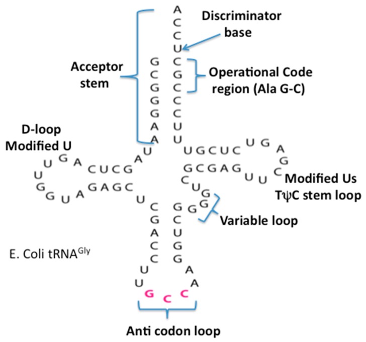
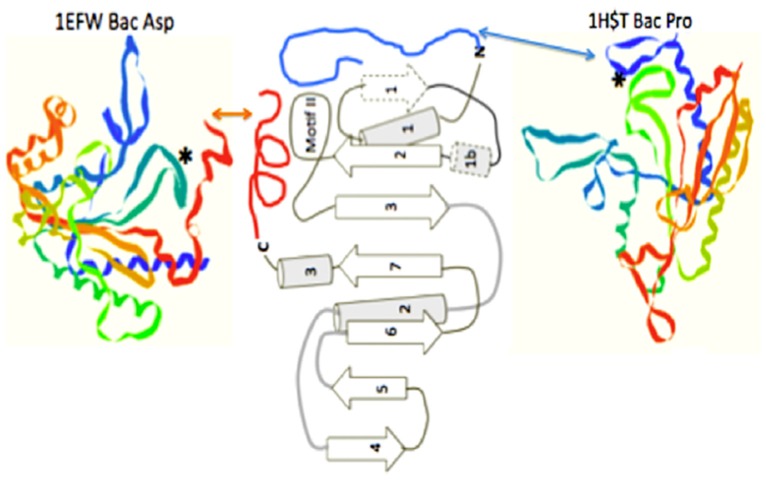
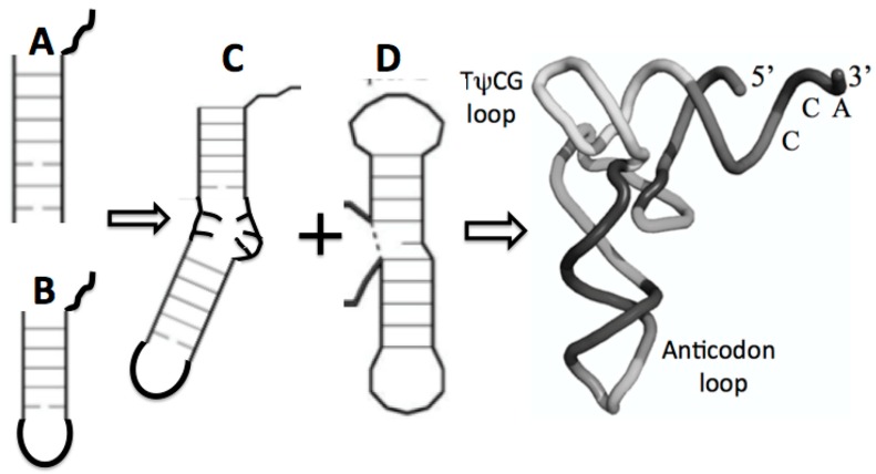
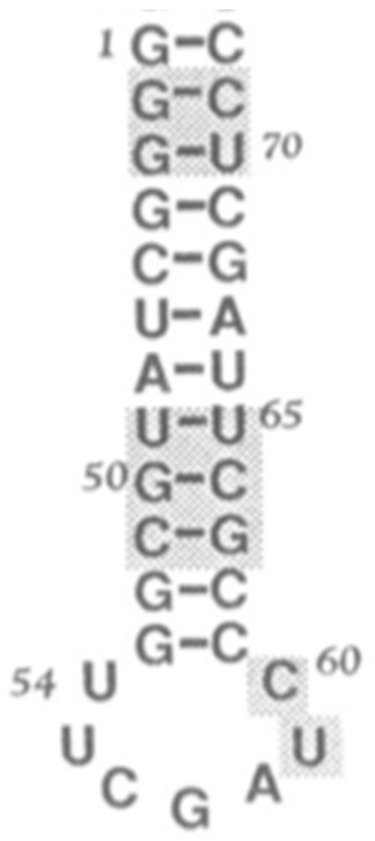
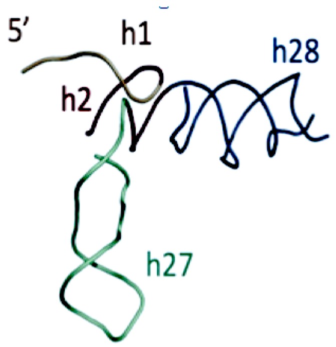
The early metabolism arising in a Thioester world gave rise to amino acids and their simple peptides. The catalytic activity of these early simple peptides became instrumental in the transition from Thioester World to a Phosphate World. This transition involved the appearances of sugar phosphates, nucleotides, and polynucleotides. The coupling of the amino acids and peptides to nucleotides and polynucleotides is the origin for the genetic code. Many of the key steps in this transition are seen in in the catalytic cores of the nucleotidyltransferases, the class II tRNA synthetases (aaRSs) and the CCA adding enzyme. These catalytic cores are dominated by simple beta hairpin structures formed in the Thioester World. The code evolved from a proto-tRNA a tetramer XCCA interacting with a proto-aminoacyl-tRNA synthetase (aaRS) activating Glycine and Proline, the initial expanded code is found in the acceptor arm of the tRNA, the operational code. It is the coevolution of the tRNA with the aaRSs that is at the heart of the origin and evolution of the genetic code. There is also a close relationship between the accretion models of the evolving tRNA and that of the ribosome.

Keywords: early peptides; genetic code origin; metabolism; nucleotidyltransferases; ribosome; tRNA accretion model; tRNA-synthetase; thioester.


Conflict of interest statement

The authors declare no conflicts of interest.

Figures

Figure 1
Figure 1
The basic Purine and Pyrimidine bases. The numbering is used in the text to identify the sources of their atoms in biosynthesis.
Figure 2
Figure 2
The reduced nucleotide genetic coding tables.
Figure 3
Figure 3
The Ornithine to Arginine pathway.
Figure 4
Figure 4
Comparison of the similarity of the double beta hairpin cores of (A) the bacterial CCA adding enzyme head domain and (B) the class II tRNA synthetase minimum core of the catalytic domain. The minimum catalytic domains are in dark gray. Note the beta hairpin components 3 and 2 in (B), which define the motif II loop; and 1 and 2 in (A), which contain the two Aspartic acids. The strand numbering for (B) is as in reference [11] for the entire traditionally-defined class II catalytic domain.
Figure 5
Figure 5
The secondary cloverleaf structure of E. coli’s tRNA Gly. The discriminator base, the operational code region, and the various stems and loops are labeled in their traditional manner.
Figure 6
Figure 6
The class II synthetase catalytic core showing two alternate operational code-contacting peptide extensions, one on the N-terminal, as seen in the bacterial Proline aaRS structure 1HST.pdb, and one on the C-terminal, as seen in the bacterial Aspartic acid aaRS structure, 1EFW.pdb. Figure is adapted from [11].
Figure 7
Figure 7
Possible maturation of the full tRNA: (A) arm with CCA attachment, fused with (B) stem loop with CCA attachment resulting in (C) the boomerang that then fused with (D) the dumbell resulting in the tRNA cloverleaf secondary structure in Figure 5 and the full 3D, folded tRNA structure pictured here.
Figure 8
Figure 8
Minihelix example, proposed by Tamura [34] as a seed for the ribosomal subunits.
Figure 9
Figure 9
Proposed SSU seed, domain A Gulen et al. [37]. The SSU domain A was proposed to include SSU helices H27 and H28, resulting in a structure similar to the structure of the “boomerang” tRNA intermediate proposed structure in Figure 7C. Figure 9 has been provided by Anton Petrov.

References

    1. Dyson F. ; Origins of Life. Cambridge Press; Cambridge, UK: 1999.
    1. Hartman H. Speculations on the origin and evolution of metabolism. J. Mol. Evol. 1975;4:359–370. doi: 10.1007/BF01732537. - DOI - PubMed
    1. Hartman H. Conjectures and reveries. Photosynth. Res. 1992;33:171–176. doi: 10.1007/BF00039179. - DOI - PubMed
    1. Goldford J.E., Hartman H., Smith T.F., Segre D. Remnants of an ancient metabolism without phosphate. Cell. 2017;168:1126–1134. doi: 10.1016/j.cell.2017.02.001. - DOI - PubMed
    1. Dorr M., Kassbohrer J., Grunert R., Kreisel G., Brand W.A., Werner R.A., Geilmann H., Apfel C., Robl C., Weigand W. A Possible prebiotic formation of ammonia from dinitrogen on iron sulfide surfaces. Angew. Chem. Int. Ed. 2003;42:1540–1543. doi: 10.1002/anie.200250371. - DOI - PubMed

LinkOut - more resources