Review
. 2019 Aug 22;8(9):333.
doi: 10.3390/antiox8090333.

Anthocyanins and Their Metabolites as Therapeutic Agents for Neurodegenerative Disease

Affiliations
Review

Anthocyanins and Their Metabolites as Therapeutic Agents for Neurodegenerative Disease

Aimee N Winter et al. Antioxidants (Basel). .

Abstract

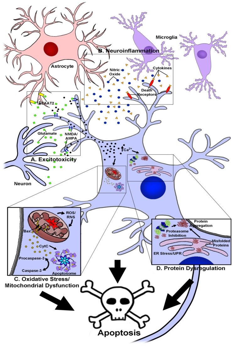
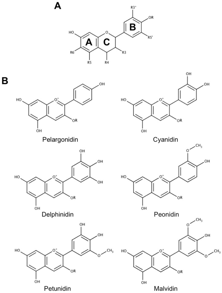
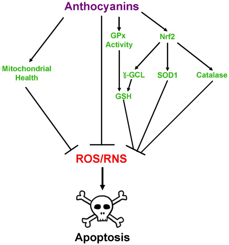
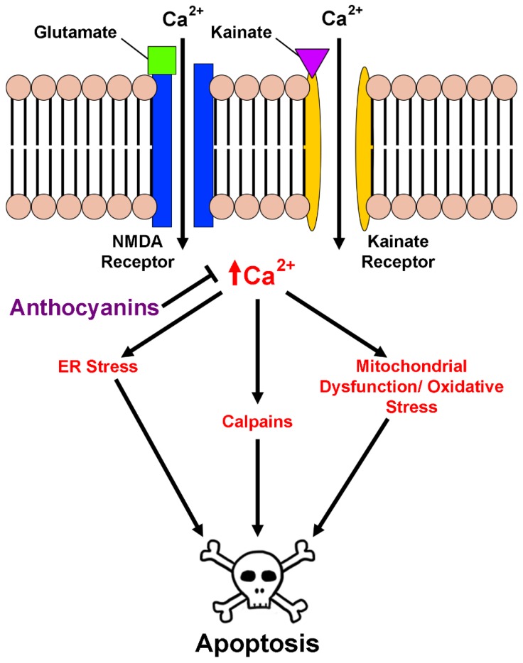
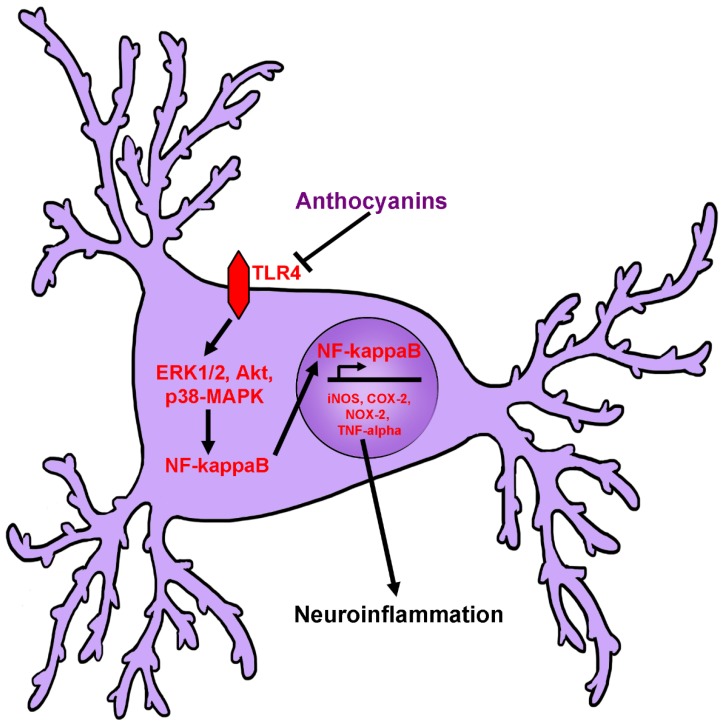
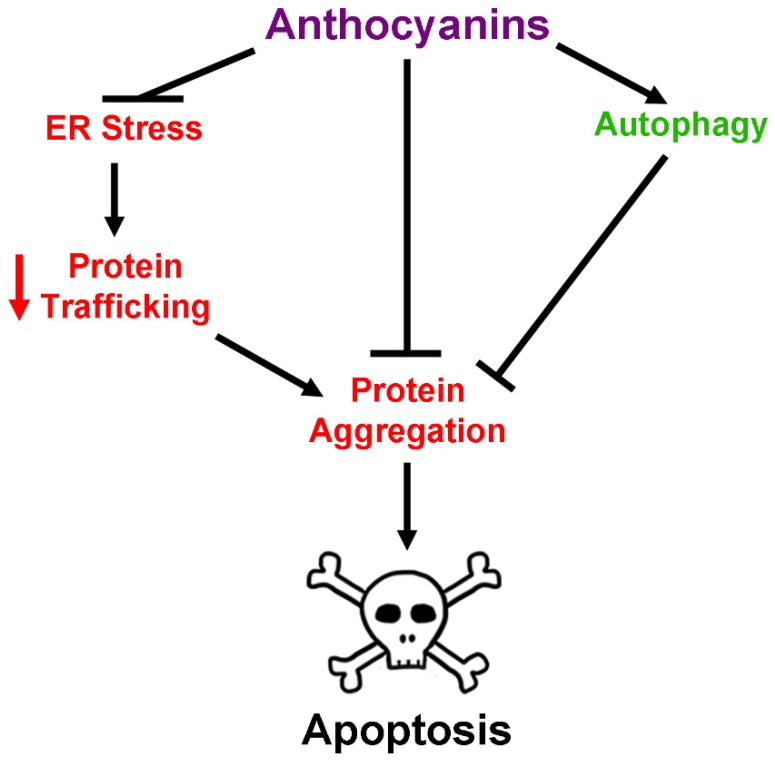
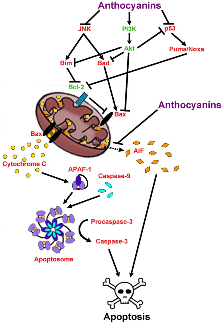
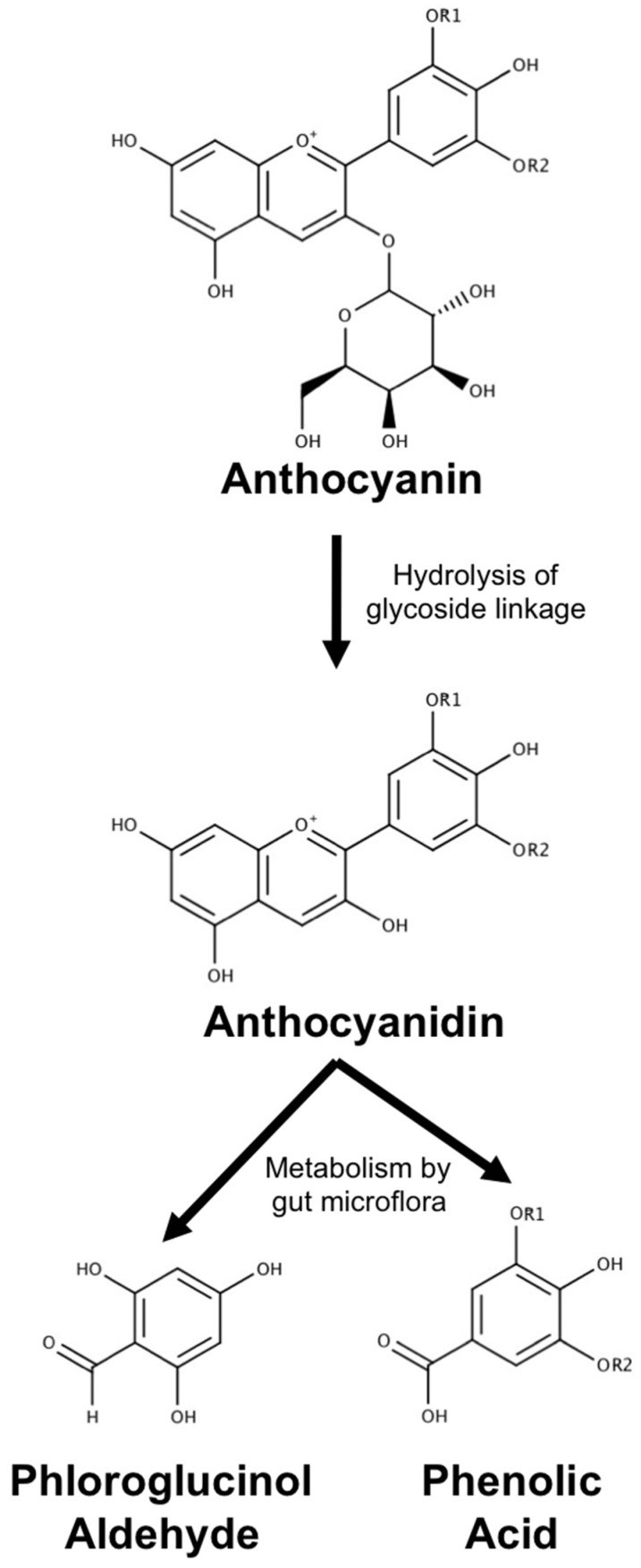
Neurodegenerative diseases, including Alzheimer's disease, Parkinson's disease, and amyotrophic lateral sclerosis (ALS), are characterized by the death of neurons within specific regions of the brain or spinal cord. While the etiology of many neurodegenerative diseases remains elusive, several factors are thought to contribute to the neurodegenerative process, such as oxidative and nitrosative stress, excitotoxicity, endoplasmic reticulum stress, protein aggregation, and neuroinflammation. These processes culminate in the death of vulnerable neuronal populations, which manifests symptomatically as cognitive and/or motor impairments. Until recently, most treatments for these disorders have targeted single aspects of disease pathology; however, this strategy has proved largely ineffective, and focus has now turned towards therapeutics which target multiple aspects underlying neurodegeneration. Anthocyanins are unique flavonoid compounds that have been shown to modulate several of the factors contributing to neuronal death, and interest in their use as therapeutics for neurodegeneration has grown in recent years. Additionally, due to observations that the bioavailability of anthocyanins is low relative to that of their metabolites, it has been proposed that anthocyanin metabolites may play a significant part in mediating the beneficial effects of an anthocyanin-rich diet. Thus, in this review, we will explore the evidence evaluating the neuroprotective and therapeutic potential of anthocyanins and their common metabolites for treating neurodegenerative diseases.

Keywords: Alzheimer’s disease; Parkinson’s disease; amyotrophic lateral sclerosis; anthocyanins; flavonoids; inflammation; neurodegeneration; neuroprotection; oxidative stress; phenolic acids.


Conflict of interest statement

The authors declare no conflict of interest.

Figures

Figure 1
Figure 1
Molecular mechanisms contributing to the pathogenesis of neurodegenerative diseases. A, The role of excitotoxicity in neurodegeneration. Glutamate is released from pre-synaptic neuron terminals in elevated quantities and binds to glutamate receptors such as N-methyl-d-aspartate (NMDA) and α-amino-3-hydroxy-5-methyl-4-isoxazolepropionic acid (AMPA) receptors. Receptor binding causes massive calcium influx in post-synaptic neurons and activates pro-apoptotic signaling cascades, in addition to inducing mitochondrial dysfunction and endoplasmic reticulum (ER) stress responses. In some diseases, such as ALS, glutamate uptake from the synapse by the excitatory amino acid transporter 2 (EAAT2) in astrocytes is impaired, exacerbating glutamate excitotoxicity. B, The role of neuroinflammation in neurodegeneration. Glial cells such as astrocytes and microglia become chronically enflamed in disease states and secrete oxidative species, such as nitric oxide, and pro-inflammatory cytokines. Cytokines bind to death receptors on the cell surface and activate pro-apoptotic signaling cascades. C, The role of oxidative stress and mitochondrial dysfunction in neurodegeneration. Mitochondrial dysfunction occurs as a result of several factors, causing mitochondria to produce elevated levels of reactive oxygen and nitrogen species (ROS and RNS). Enhanced production of ROS and RNS exacerbates mitochondrial dysfunction, eventually causing release of the pro-apoptotic signaling protein, cytochrome c (CytC). Cytochrome C contributes to formation of the apoptosome, which in turn cleaves procaspase-3 to form active caspase-3, stimulating apoptosis. D, The role of protein dysregulation in neurodegeneration. ER stress occurs as a result of multiple factors, such as the presence of mutated or damaged proteins, causing accumulation of misfolded proteins and activation of the unfolded protein response (UPR). Misfolded or mutated proteins are trafficked to the proteasome for degradation. As these proteins accumulate and aggregate, the proteasome becomes clogged, leading to proteasome inhibition and further accumulation of protein aggregates. Protein aggregation and ER stress trigger pro-apoptotic signaling cascades. Collectively, these factors lead to death of vulnerable neuronal populations.
Figure 2
Figure 2
Common anthocyanin structures. (A) General flavonoid structure. Flavonoids possess a characteristic three-ring structure that is conserved across all family members. Several classes of flavonoids exist, including anthocyanins, which differ depending on substitutions of the A, B, and C-rings. (B) Structures of the six most common anthocyanins. Anthocyanins possess a cationic structure that differs between species predominately in substitutions of the B-ring. Anthocyanins also possess a sugar moiety as a part of their structure, represented as R. Common sugar moieties include but are not limited to glucose, galactose, and rutinose. All structures included in this review were created using MarvinSketch (ChemAxon, Cambridge, MA 02138, USA).
Figure 3
Figure 3
Antioxidant effects of anthoycanins. Anthocyanins modulate damage produced by reactive oxygen and nitrogen species (ROS and RNS) by several mechanisms. These include direct enhancement of glutathione peroxidase (GPx) activity, direct scavenging of ROS and RNS, activation of nuclear factor erythroid 2-related factor 2 (Nrf-2) transcription of antioxidant enzymes, and promotion of mitochondrial health and function. Genes activated by Nrf2 include, but are not limited to, those for catalase, Cu,Zn-superoxide dismutase (SOD1), and gamma-glutamylcysteine ligase (γ-GCL), which increases synthesis of the critical antioxidant, glutathione (GSH). GSH can then be used in conjunction with GPx and another enzyme, glutathione reductase, to scavenge oxidative species. Collectively, these mechanisms detoxify ROS and RNS to prevent apoptosis.
Figure 4
Figure 4
Effects of anthocyanins on calcium homeostasis and excitotoxicity. Binding of the excitatory compounds, glutamate or kainate, to their cognate receptors on the cell membrane causes massive calcium influx into neurons. This calcium influx interferes with the protein folding functions of the ER, resulting in ER stress, activation of the UPR, and subsequent activation of pro-apoptotic signaling cascades if not resolved. Additionally, high levels of intracellular calcium cause membrane depolarization at the mitochondria and uncoupling of the electron transport chain, leading to mitochondrial dysfunction, oxidative stress, opening of the permeability transition pore and release of apoptogenic factors into the cytosol. High calcium concentrations can also lead to direct activation of pro-apoptotic factors such as calpains, leading to cell death. Anthocyanins protect neurons from excitotoxicity by preventing increases in intracellular calcium caused by glutamate and kainate signaling.
Figure 5
Figure 5
Effects of anthocyanins on neuroinflammation. Inflammatory stimuli, such as deposits of aggregated proteins, cause activation of toll-like receptor-4 (TLR4), and downstream induction of extracellular regulated signal kinase 1/2 (ERK1/2), Akt, and p38-mitogen-activated protein kinase (p38-MAPK), which subsequently activate nuclear factor-κB (NF-κB) in microglia and astrocytes. NF-κB then translocates to the nucleus and initiates transcription of pro-inflammatory genes including, but not limited to inducible nitric oxide synthase (iNOS), cyclooxygenase-2 (COX-2) NADPH-oxidase-2 (NOX-2), and tumor necrosis factor-α (TNF-α). It is thought that anthocyanins inhibit this pathway by blocking activation of TLR4, ERK1/2, Akt, and p38-MAPK.
Figure 6
Figure 6
Effects of anthocyanins on protein dysregulation and homeostasis. Protein homeostasis is disrupted in several ways in neurodegenerative disease. Increased levels of ER stress cause significant decreases in protein trafficking to other organelles such as the Golgi apparatus. Decreased protein trafficking results in accumulation of misfolded and mutant proteins, causing protein aggregates to form. Formation of these toxic aggregates then contributes to induction of neuronal apoptosis. Anthocyanins modulate this process by reducing ER stress, directly inhibiting the formation of toxic protein aggregates, and stimulating autophagy processes to clear aggregates formed within the neuron.
Figure 7
Figure 7
Effects of anthocyanins on pro-survival and pro-apoptotic signaling pathways. Anthocyanins modulate several signaling pathways involved in cell survival and death. Anthocyanins inhibit the activity of c-Jun N-terminal kinase (JNK) and p53, which are responsible for activating pro-apoptotic family members of the Bcl-2 family of proteins, Bim, Bad, Puma, and Noxa. Bim, Puma, and Noxa are known to inhibit the pro-survival functions of B-cell lymphoma-2 (Bcl-2), causing activation of the pro-apoptotic protein, Bax. Bax can also be activated by interaction with Bad. Bax then forms pores in the mitochondrial membrane, allowing the release of cytochrome c from mitochondria. Cytochrome c interacts with apoptosis protease activating factor-1 (APAF-1) and caspase-9 to form the apoptosome. The apoptosome then cleaves procaspase-3 to form active caspase-3, stimulating apoptosis. Anthocyanins also enhance the activity of the phosphoinositide-3-kinase (PI3K)/Akt pro-survival signaling pathway, which inhibits activity of pro-apoptotic Bcl-2 family members including Bim, Bad, and Bax, in addition to inhibiting the activity of p53. This activity inhibits entry of neurons into caspase-dependent apoptosis. Alternatively, anthocyanins have also been shown to inhibit caspase-independent apoptosis by blocking the translocation of apoptosis inducing factor (AIF) from mitochondria to the cytosol and subsequently, the nucleus (dashed arrow).
Figure 8
Figure 8
Metabolism of anthocyanins. A generic anthocyanin with a glucoside moiety is pictured. Parent anthocyanin species are first converted to an aglycon (anthocyanidin) form by hydrolysis of glycoside linkages in the small intestine. Upon entry into the large intestine, the anthocyanidin is further metabolized by gut microflora to produce a universal aldehyde metabolite, phloroglucinol aldehyde, and a phenolic acid that retains the structure of the B-ring of the parent anthocyanin.
Figure 9
Figure 9
Common phenolic acid metabolites derived from anthocyanins. (A) Common anthocyanin bases and their phenolic acid metabolites. (B) Molecular structures of six common phenolic acid metabolites derived from anthocyanins.
Figure 10
Figure 10
The neuroprotective activities of anthocyanins metabolites. The phenolic acid metabolites display a host of neuroprotective activities. These compounds have been shown to have potent antioxidant abilities through the activation of antioxidant enzymes and direct scavenging of ROS, and are known to prevent excitotoxicity by preserving calcium homeostasis. Additionally, phenolic acids prevent neuroinflammation by reducing the expression of pro-inflammatory pathways in astrocytes and microglia. Many phenolic acids have been shown to directly interfere with aggregation of proteins such as amyloid beta and alpha-synuclein. Moreover, the phenolic acid metabolites of anthocyanins activate pro-survival signaling pathways, while inhibiting expression and activation of pro-apoptotic signaling cascades. Uniquely, these compounds have also been shown to promote neurite outgrowth and axonal health, which may preserve important signaling networks in the brain and spinal cord.

References

    1. Ilieva H., Polymenidou M., Cleveland D.W. Non-cell autonomous toxicity in neurodegenerative disorders: ALS and beyond. J. Cell Biol. 2009;187:761–772. doi: 10.1083/jcb.200908164. - DOI - PMC - PubMed
    1. Taylor J.P., Brown R.H., Jr., Cleveland D.W. Decoding ALS: From genes to mechanism. Nature. 2016;539:197–206. doi: 10.1038/nature20413. - DOI - PMC - PubMed
    1. Magalingam K.B., Radhakrishnan A., Ping N.S., Haleagrahara N. Current Concepts of Neurodegenerative Mechanisms in Alzheimer’s Disease. Biomed. Res. Int. 2018;2018:3740461. doi: 10.1155/2018/3740461. - DOI - PMC - PubMed
    1. Zeng X.S., Geng W.S., Jia J.J., Chen L., Zhang P.P. Cellular and Molecular Basis of Neurodegeneration in Parkinson Disease. Front. Aging Neurosci. 2018;10:109. doi: 10.3389/fnagi.2018.00109. - DOI - PMC - PubMed
    1. Wallace T.C. Anthocyanins in cardiovascular disease. Adv. Nutr. 2011;2:1–7. doi: 10.3945/an.110.000042. - DOI - PMC - PubMed

LinkOut - more resources