. 2019 Dec;138(6):885-900.
doi: 10.1007/s00401-019-02061-5. Epub 2019 Aug 23.

Dissecting the genetic basis of focal cortical dysplasia: a large cohort study

Affiliations

Dissecting the genetic basis of focal cortical dysplasia: a large cohort study

Sara Baldassari et al. Acta Neuropathol. 2019 Dec.

Abstract

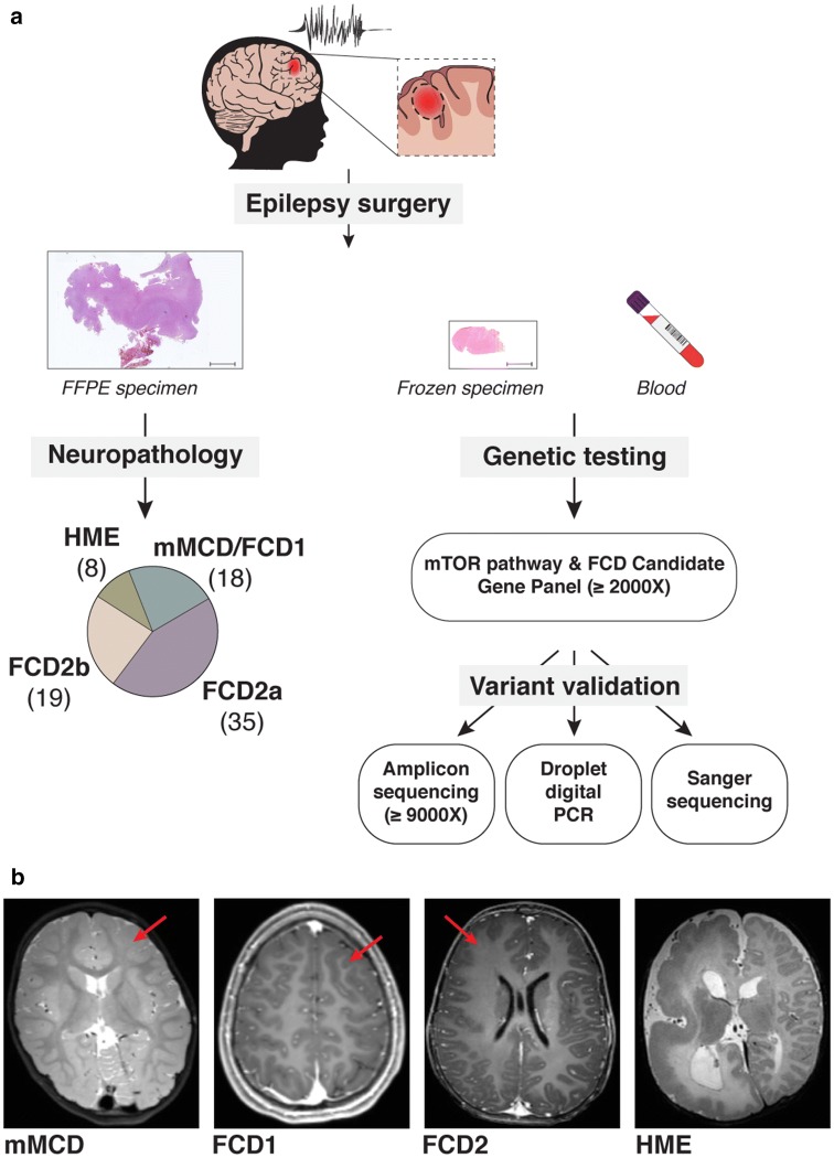
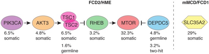
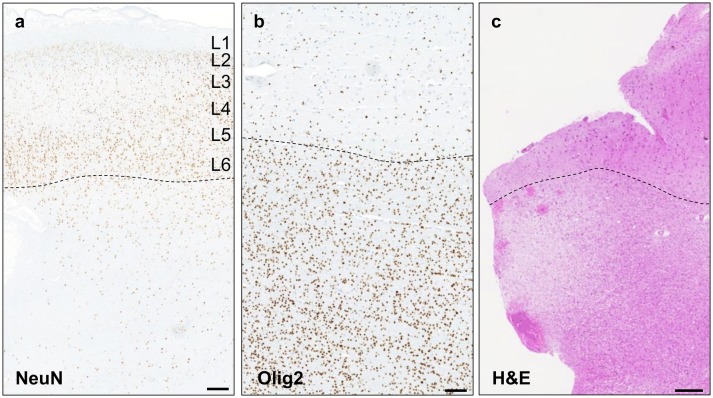
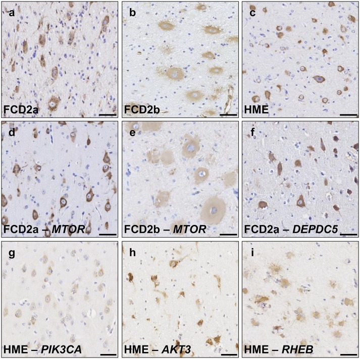
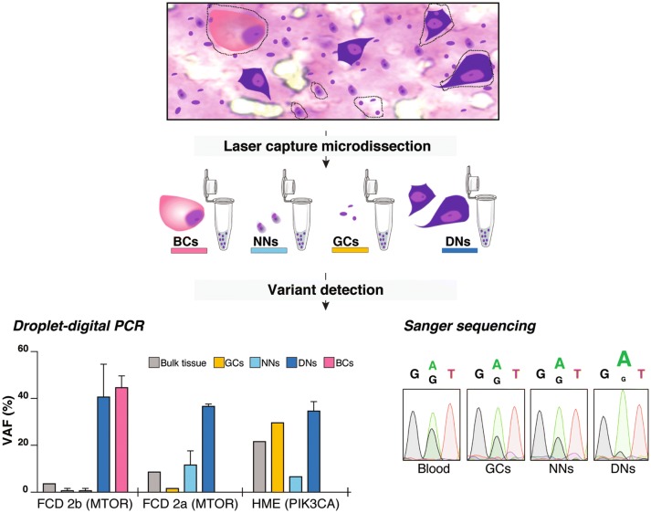
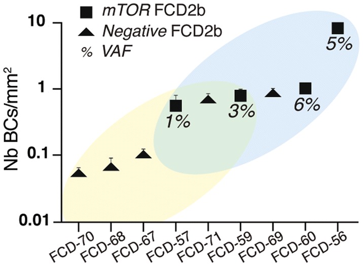
Genetic malformations of cortical development (MCDs), such as mild MCDs (mMCD), focal cortical dysplasia (FCD), and hemimegalencephaly (HME), are major causes of severe pediatric refractory epilepsies subjected to neurosurgery. FCD2 are characterized by neuropathological hallmarks that include enlarged dysmorphic neurons (DNs) and balloon cells (BCs). Here, we provide a comprehensive assessment of the contribution of germline and somatic variants in a large cohort of surgical MCD cases. We enrolled in a monocentric study 80 children with drug-resistant epilepsy and a postsurgical neuropathological diagnosis of mMCD, FCD1, FCD2, or HME. We performed targeted gene sequencing ( ≥ 2000X read depth) on matched blood-brain samples to search for low-allele frequency variants in mTOR pathway and FCD genes. We were able to elucidate 29% of mMCD/FCD1 patients and 63% of FCD2/HME patients. Somatic loss-of-function variants in the N-glycosylation pathway-associated SLC35A2 gene were found in mMCD/FCD1 cases. Somatic gain-of-function variants in MTOR and its activators (AKT3, PIK3CA, RHEB), as well as germline, somatic and two-hit loss-of-function variants in its repressors (DEPDC5, TSC1, TSC2) were found exclusively in FCD2/HME cases. We show that panel-negative FCD2 cases display strong pS6-immunostaining, stressing that all FCD2 are mTORopathies. Analysis of microdissected cells demonstrated that DNs and BCs carry the pathogenic variants. We further observed a correlation between the density of pathological cells and the variant-detection likelihood. Single-cell microdissection followed by sequencing of enriched pools of DNs unveiled a somatic second-hit loss-of-heterozygosity in a DEPDC5 germline case. In conclusion, this study indicates that mMCD/FCD1 and FCD2/HME are two distinct genetic entities: while all FCD2/HME are mosaic mTORopathies, mMCD/FCD1 are not caused by mTOR-pathway-hyperactivating variants, and ~ 30% of the cases are related to glycosylation defects. We provide a framework for efficient genetic testing in FCD/HME, linking neuropathology to genetic findings and emphasizing the usefulness of molecular evaluation in the pediatric epileptic neurosurgical population.

Keywords: Brain mosaicism; Epilepsy-associated focal cortical dysplasia; Neurogenetics; Somatic variant; mTOR pathway.


Conflict of interest statement

The authors have declared that no conflict of interest exists.

Figures

Fig. 1
Fig. 1
Study workflow. a The study includes children who underwent epilepsy surgery with suspected cortical brain malformation of development, including mMCD, FCD (types 1 and 2), or HME. For each patient, we collected both FFPE (formalin-fixed paraffin-embedded tissue) and fresh frozen brain specimens, and matched blood samples. Neuropathological examination allowed classification of mild MCD (mMCD), FCD1, FCD2a, FCD2b, and HME subtypes in 80 samples. Targeted capture-based panel sequencing of mTOR pathway and FCD candidate genes revealed germline, somatic, and two-hits variants, which were subsequently confirmed by alternative sequencing methods (amplicon or conventional Sanger) or droplet digital PCR. b Representative 3 T MRI images are shown for mMCD (FCD-1 case), FCD1 (FCD-9 case), FCD2 (FCD-30 case) and for HME (HME-76 case)
Fig. 2
Fig. 2
Overall genetics findings. Global diagnostic yield among mMCD/FCD1, FCD2, and HME cases are illustrated. The percentage of patients carrying pathogenic variants among the panel-positive cases (n = 43) is indicated to denote the contribution of each gene to the pathogenesis of mMCD/FCD/HME
Fig. 3
Fig. 3
Genotype–phenotype correlations. Comparison of age at seizure (sz) onset between groups of patients with variants in different genes, reported as boxplots with minimum, maximum, average, and standard deviation (Kruskal–Wallis test with multiple comparisons). Germline group includes five DEPDC5 germline cases and one TSC2 germline case. TSC1/2 group includes only somatic variants
Fig. 4
Fig. 4
Histological findings in SLC35A2-mMCD2 brain specimens. a NeuN immunoreactivity of the middle frontal cortex region from patient FCD-2 shows blurred grey–white matter border (dashed line) and excess of heterotopic neurons ( > 30/mm2); scale bar: 1 mm. b Increased oligodendrocyte density in the white matter (below the dashed line) in the fronto-pre-central brain tissue from patient FCD-4; scale bar: 500 µm. c H&E staining of the pre-motor cortex tissue in patient FCD-4 shows white matter pallor; scale bar: 250 µm
Fig. 5
Fig. 5
Activation of the mTOR pathway in FCD2/HME cases. pS6 (pS6 Ser240/244) immunostaining on 4 μm-FFPE brain sections in panel-negative (ac) and panel-positive FCD2/HME cases (di). Strong pS6 immunoreactivity in DNs and BCs was detected in all cases, though signals appeared less intense in BCs. Panel-negative representative cases are shown to illustrate: FCD2a in a (FCD-40), FCD2b in b (FCD-68) and HME in c (HME-80). Panel-positive representative cases: FCD2a MTOR:p.Ser2215Phe in d (FCD-24), FCD2b MTOR:p.Thr1977Lys in e (FCD-56), FCD2a DEPDC5:p.Arg239* in f (FCD-35). HME cases with variants in different genes are reported in panels g–i: PIK3CA:p.Glu542Lys in g (HME-75), AKT3:p.Glu17Lys in h (HME-74); RHEB:Tyr35Leu in i (HME-79). Scale bar: 50 µm
Fig. 6
Fig. 6
Balloon cells and dysmorphic neurons carry pathogenic variants. Characterization of variant-carrying cells and LOH evidence by laser-capture microdissection. DNs, BCs, neurons with normal appearance (NNs) and glial cells (GCs) were laser-captured and pooled for DNA extraction. Left panel: For MTOR and PIK3CA variants detection, we performed droplet digital PCR on DNA extracted from various pools: 20–100 cells for DNs, two pools of 15 and 22 cells for BCs, pools of 50–100 cells for NNs and pools of 100–200 cells for GCs. The histogram illustrates the variant allele frequency (VAF, in percentage) detected by droplet digital PCR (ddPCR) in three patients (FCD-25, FCD-56 and HME-77). A single ddPCR experiment was performed on bulk DNAs and for laser-captured NNs and GCs in FCD2a and HME samples. For all laser-captured cell types in FCD2b sample and for laser-captured DNs in all samples, the ddPCR experiment was performed in duplicate to achieve significance: mean variant allele frequency values and standard error of the means are reported in these cases. Right panel: Sanger sequencing on DNA extracted from pools of 250 DNs, 300 NNs and 300 GCs was performed to confirm DEPDC5 somatic LOH in patient FCD-36
Fig. 7
Fig. 7
Balloon cells density correlates with genetic findings. Balloon cells density (number of BC/mm2) in five panel-negative (yellow region in the plot) and four panel-positive (blue region in the plot) FCD2b cases. The average density calculated on three sections per each patient and the standard error of the mean is reported. The two groups overlap at intermediate density values (green region in the plot)

References

    1. Abdijadid S, Mathern GW, Levine MS, Cepeda C. Basic mechanisms of epileptogenesis in pediatric cortical dysplasia. CNS Neurosci Ther. 2015;21:92–103. - PMC - PubMed
    1. Baek ST, Copeland B, Yun EJ, Kwon SK, Guemez-Gamboa A, Schaffer AE, et al. An AKT3-FOXG1-reelin network underlies defective migration in human focal malformations of cortical development. Nat Med. 2015;21:1445–1454. - PMC - PubMed
    1. Baldassari S, Picard F, Verbeek NE, van Kempen M, Brilstra EH, Lesca G, et al. The landscape of epilepsy-related GATOR1 variants. Genet Med. 2019;21:398–408. - PMC - PubMed
    1. Barkovich AJ, Dobyns WB, Guerrini R. Malformations of cortical development and epilepsy. Cold Spring Harb Perspect Med. 2015;5:a022392. - PMC - PubMed
    1. Baulac S, Ishida S, Marsan E, Miquel C, Biraben A, Nguyen DK, et al. Familial focal epilepsy with focal cortical dysplasia due to DEPDC5 mutations. Ann Neurol. 2015;77:675–683. - PubMed

Publication types