

Deep Learning Classifiers for Automated Detection of Gonioscopic Angle Closure Based on Anterior Segment OCT Images
- PMID: 31445003
- PMCID: PMC6888901
- DOI: 10.1016/j.ajo.2019.08.004
Deep Learning Classifiers for Automated Detection of Gonioscopic Angle Closure Based on Anterior Segment OCT Images
Abstract
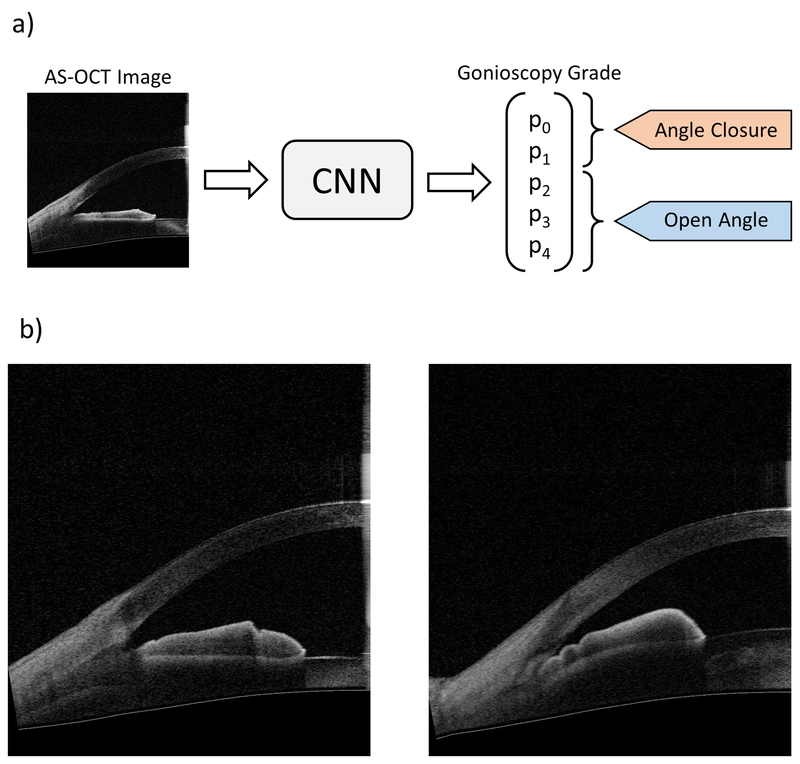
Purpose: To develop and test deep learning classifiers that detect gonioscopic angle closure and primary angle closure disease (PACD) based on fully automated analysis of anterior segment OCT (AS-OCT) images.
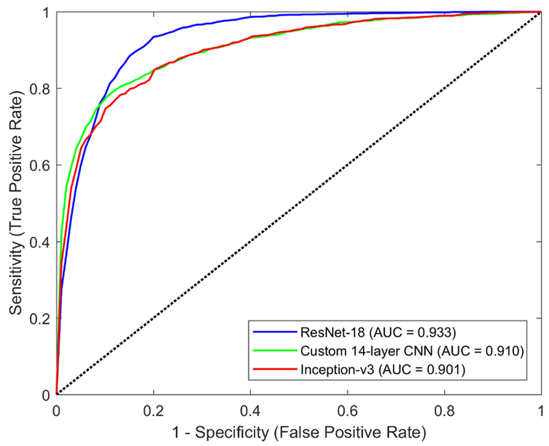
Methods: Subjects were recruited as part of the Chinese-American Eye Study (CHES), a population-based study of Chinese Americans in Los Angeles, California, USA. Each subject underwent a complete ocular examination including gonioscopy and AS-OCT imaging in each quadrant of the anterior chamber angle (ACA). Deep learning methods were used to develop 3 competing multi-class convolutional neural network (CNN) classifiers for modified Shaffer grades 0, 1, 2, 3, and 4. Binary probabilities for closed (grades 0 and 1) and open (grades 2, 3, and 4) angles were calculated by summing over the corresponding grades. Classifier performance was evaluated by 5-fold cross-validation and on an independent test dataset. Outcome measures included area under the receiver operating characteristic curve (AUC) for detecting gonioscopic angle closure and PACD, defined as either 2 or 3 quadrants of gonioscopic angle closure per eye.
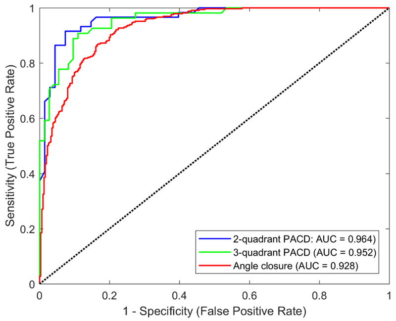
Results: A total of 4036 AS-OCT images with corresponding gonioscopy grades (1943 open, 2093 closed) were obtained from 791 CHES subjects. Three competing CNN classifiers were developed with a cross-validation dataset of 3396 images (1632 open, 1764 closed) from 664 subjects. The remaining 640 images (311 open, 329 closed) from 127 subjects were segregated into a test dataset. The best-performing classifier was developed by applying transfer learning to the ResNet-18 architecture. For detecting gonioscopic angle closure, this classifier achieved an AUC of 0.933 (95% confidence interval, 0.925-0.941) on the cross-validation dataset and 0.928 on the test dataset. For detecting PACD based on 2- and 3-quadrant definitions, the ResNet-18 classifier achieved AUCs of 0.964 and 0.952, respectively, on the test dataset.
Conclusion: Deep learning classifiers effectively detect gonioscopic angle closure and PACD based on automated analysis of AS-OCT images. These methods could be used to automate clinical evaluations of the ACA and improve access to eye care in high-risk populations.
Copyright © 2019 Elsevier Inc. All rights reserved.
Figures
References
-
- Tham YC, Li X, Wong TY, Quigley HA, Aung T, Cheng CY. Global prevalence of glaucoma and projections of glaucoma burden through 2040: A systematic review and meta-analysis. Ophthalmology. 2014;121(11):2081–2090. - PubMed
-
- Liang Y, Friedman DS, Zhou Q, et al. Prevalence and characteristics of primary angle-closure diseases in a rural adult Chinese population: The Handan eye study. Investig Ophthalmol Vis Sci. 2011;52(12):8672–8679. - PubMed
-
- Sawaguchi S, Sakai H, Iwase A, et al. Prevalence of primary angle closure and primary angle-closure glaucoma in a southwestern rural population of Japan: The Kumejima study. Ophthalmology. 2012;119(6): 1134–1142. - PubMed
-
- He M, Friedman DS, Ge J, et al. Laser Peripheral Iridotomy in Primary Angle-Closure Suspects: Biometric and Gonioscopic Outcomes. The Liwan Eye Study. Ophthalmology. 2007;114(3):494–500. - PubMed
-
- Azuara-Blanco A, Burr J, Ramsay C, et al. Effectiveness of early lens extraction for the treatment of primary angle-closure glaucoma (EAGLE): a randomised controlled trial. Lancet. 2016;388(10052): 1389–1397. - PubMed
Publication types
MeSH terms
Grants and funding
LinkOut - more resources
Full Text Sources
Other Literature Sources
Research Materials
