. 2019 Oct 1:97:272-280.
doi: 10.1016/j.actbio.2019.08.031. Epub 2019 Aug 22.

A hydrogel platform for in vitro three dimensional assembly of human stem cell-derived islet cells and endothelial cells

Affiliations

A hydrogel platform for in vitro three dimensional assembly of human stem cell-derived islet cells and endothelial cells

Punn Augsornworawat et al. Acta Biomater. .

Abstract

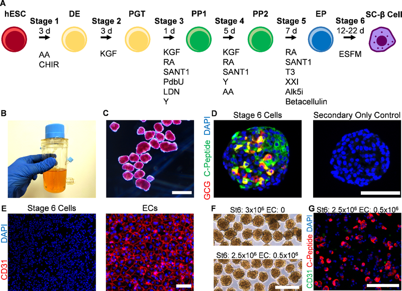
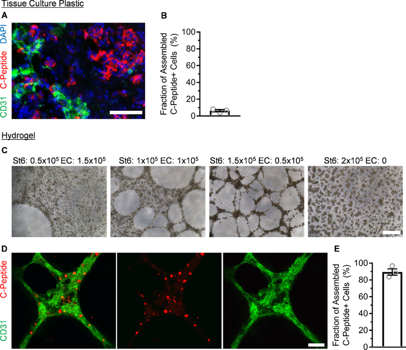
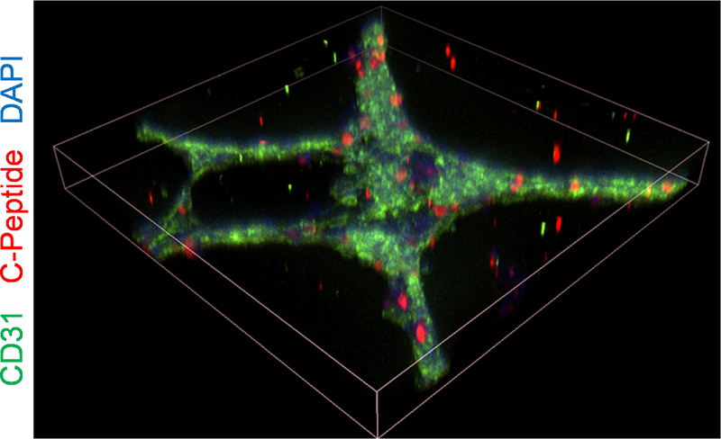
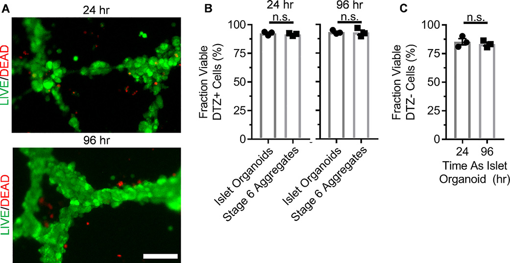
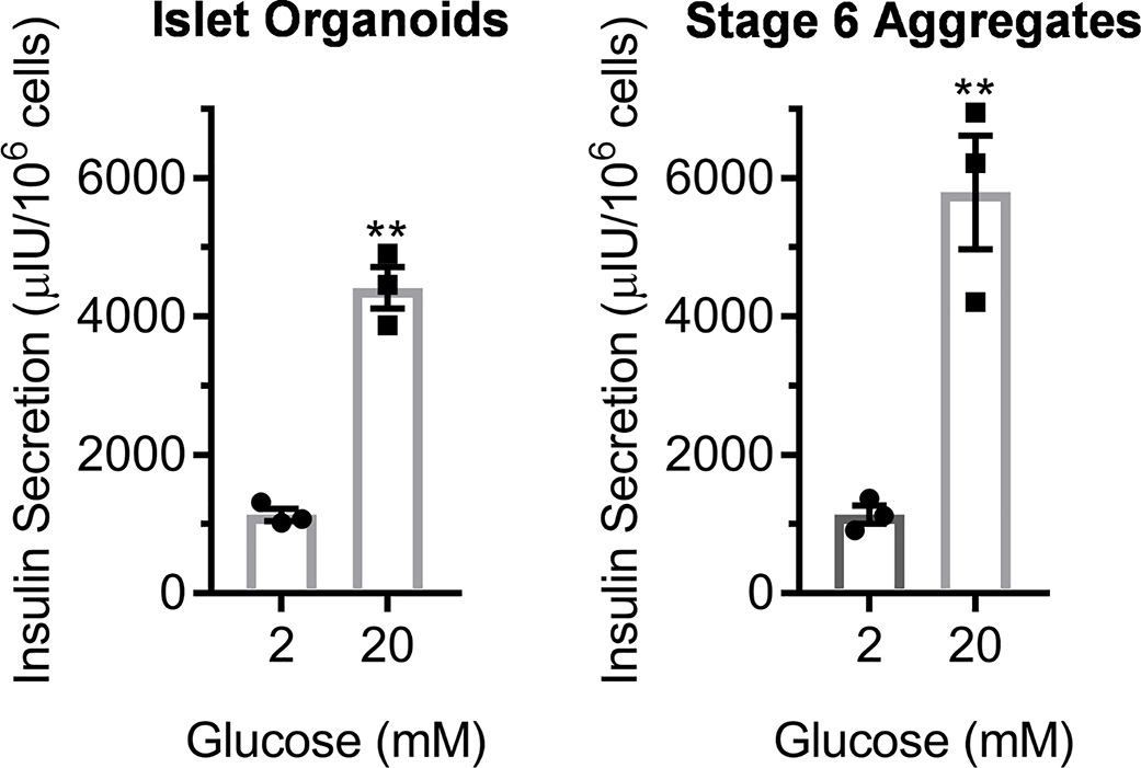
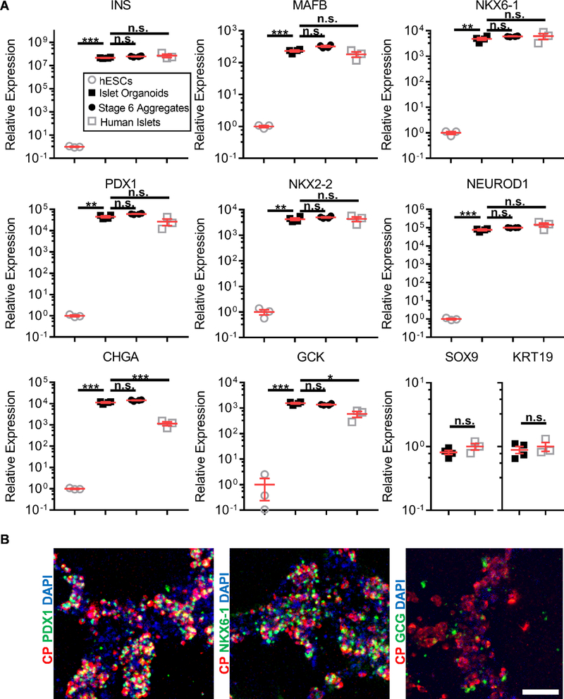
Differentiation of stem cells into functional replacement cells and tissues is a major goal of the regenerative medicine field. However, one limitation has been organization of differentiated cells into multi-cellular, three-dimensional assemblies. The islets of Langerhans contain many endocrine and non-endocrine cell types, such as insulin-producing β cells and endothelial cells. Despite the potential importance of endothelial cells to islet function, facilitating interactions between endothelial cells and islet endocrine cell types already differentiated from human embryonic stem cells has been difficult in vitro. We have developed a strategy of assembling human embryonic stem cell-derived islet cells with endothelial cells into three-dimensional aggregates on a hydrogel. The resulting islet organoids express β cell and other endocrine markers and are functional, capable of undergoing glucose-stimulated insulin secretion. This assembly was not observed on traditional tissue culture plastic and in aggregates generated in suspension culture, highlighting how physical culture conditions greatly influence the interactions among these cell types. These results provide a platform for evaluating the effects of the islet tissue microenvironment on human embryonic stem cell-derived β cells and other islet endocrine cells to develop tissue engineered islets. STATEMENT OF SIGNIFICANCE: Differentiation of insulin-producing cells and tissues from human pluripotent stem cells is being investigated for diabetes cell replacement therapies. Despite successes generating β cells, the cell type responsible for glucose-stimulated insulin secretion within the islets of Langerhans found in the pancreas, successful assembly with other non-endocrine cell types, particularly endothelial cells, has been technically challenging. The present study provides a platform for the assembly of endothelial cells with SC-β and other endocrine cells, producing islet organoids that are functional and express β cell markers, that can be used to study the islet microenvironment and islet tissue engineering.

Keywords: Biomaterials; Diabetes; Organoids; Stem cells; Tissue engineering.


Figures

Fig. 1.
Fig. 1.
Generation of SC-β cell aggregates. A. Schematic diagram of differentiation process, illustrating cell fate changes to generate SC-β cells. The entire procedure is done in cellular aggregates. B. Image of spinner flask approach used to grow and differentiate hESCs to SC-β cells. C. Micrograph of Stage 6 clusters stained with dithizone, a dye that stains β cells red, imaged under bright field. Scale bar = 500 μm. D. Immunostaining of Stage 6 cluster sectioned and stained for C-peptide, which is produced by β cells, and glucagon, which is produced by α cells. Both primary and secondary antibodies were used for the image on the left but only secondary antibodies for the image on the right. These images were taken with the same settings. Scale bar = 100 μm. E. Immunostaining of dispersed Stage 6 clusters (left) and ECs (right) plated for assessment for CD31, an EC marker. These images were taken with the same settings. Scale bar = 100 μm. F. Micrographs of unstained reaggregated Stage 6 clusters with or without the addition of ECs after 24 hr. Scale bar = 400 μm. G. Immunostaining of Stage 6 clusters reaggregated with ECs after 24 hr then dispersed and plated 24 hr for assessment. Scale bar = 150 μm. DE, definitive endoderm; PGT, primitive gut tube; PP1, pancreatic progenitor 1; PP2, pancreatic progenitor 2; EP, endocrine progenitor; AA, activin A; CHIR, CHIR9901; KGF, keratinocyte growth factor; RA, retinoic acid; Y, Y27632; LDN, LDN193189; PdbU, phorbol 12,13-dibutyrate; T3, triiodothyronine; Alk5i, Alk5 inhibitor type II; ESFM, enriched serum-free medium.
Fig. 2.
Fig. 2.
ECs and SC-β cells assemble on top of Matrigel hydrogel but not tissue culture plastic. A. Immunostaining of Stage 6 clusters mixed with ECs and plated on tissue culture plastic for assembly after 24 hr. Scale bar = 150 μm. B. Quantification of the fraction of C-peptide+ cells in contact with CD31+ cells using the tissue culture plastic approach. n=3. C. Bright field micrographs of varying ratios of ECs and SC-β cells plated on a slab of Matrigel hydrogel for assembly after 24 hr. Scale bar = 400 μm. D. En face image of immunostained organoid produced with a 3:1 ratio of SC-β cells and ECs after 24 hr. Scale bar = 100 μm. E. Quantification of the fraction of C-peptide+ cells in contact with CD31+ cells after 24 hr using the hydrogel approach. n=3.
Fig. 3.
Fig. 3.
Confocal image construction of islet organoid produced with 3:1 ratio of SC-β cells and ECs after 24 hr. Tilted view is constructed from three-dimensional z-stacks, and the shown dimensions are 636.4 × 636.4 × 93 μm.
Fig. 4.
Fig. 4.
Viability assessment of ECs and SC-β cells assembly formed on Matrigel. A. En face image of organoids stained with viability dye indicating live (green) and dead (red) cells. B. Quantification of the fraction of DTZ+ cells, marking the SC-β cell population, that are viable as assessed by trypan blue staining. p>0.05 by two-way unpaired t-test. n=3. C. Quantification of the fraction of DTZ- cells that are viable as assessed by trypan blue staining. n.s. p>0.05 by two-way unpaired t-test. n=3.
Fig. 5.
Fig. 5.
Glucose-stimulated insulin secretion assay of EC and SC-β cell assembly formed on Matrigel. 24 hr after (left) compared to Stage 6 aggregates containing SC-β cells without ECs (right). **p<0.01 by two-way paired t-test comparing low to high glucose. n=3.
Fig. 6.
Fig. 6.
Marker analysis of EC and SC-β cell assembly formed on Matrigel. A. Ten genes associated with the β cells and the pancreas were measured, comparing the islet organoids 24 hr after (n=4) to undifferentiated hESCs (n=3), Stage 6 aggregates containing SC-β cells without ECs (n=4), and human islets (n=3). n.s. p>0.05, *p<0.05, **p<0.01, and ***p<0.001 by two-way unpaired t-test. B. En face image of immunostained organoid. CP=C-Peptide. Scale bar = 100 μm.

References

    1. Bellin MD, Barton FB, Heitman A, Harmon JV, Kandaswamy R, Balamurugan AN, Sutherland DE, Alejandro R, Hering BJ, Potent induction immunotherapy promotes long-term insulin independence after islet transplantation in type 1 diabetes, Am J Transplant 12(6) (2012) 1576–83. - PMC - PubMed
    1. McCall M, Shapiro AM, Update on islet transplantation, Cold Spring Harb Perspect Med 2(7) (2012) a007823. - PMC - PubMed
    1. Millman JR, Xie C, Van Dervort A, Gurtler M, Pagliuca FW, Melton DA, Generation of stem cell-derived beta-cells from patients with type 1 diabetes, Nat Commun 7 (2016) 11463. - PMC - PubMed
    1. Pagliuca FW, Millman JR, Gurtler M, Segel M, Van Dervort A, Ryu JH, Peterson QP, Greiner D, Melton DA, Generation of functional human pancreatic beta cells in vitro, Cell 159(2) (2014) 428–39. - PMC - PubMed
    1. Velazco-Cruz L, Song J, Maxwell KG, Goedegebuure MM, Augsornworawat P, Hogrebe NJ, Millman JR, Acquisition of Dynamic Function in Human Stem Cell-Derived beta Cells, Stem cell reports 12(2) (2019) 351–365. - PMC - PubMed

Publication types