. 2019 Aug 24;11(16):6134-6152.
doi: 10.18632/aging.102177. Epub 2019 Aug 24.

Beta-amyloid induces apoptosis of neuronal cells by inhibition of the Arg/N-end rule pathway proteolytic activity

Affiliations

Beta-amyloid induces apoptosis of neuronal cells by inhibition of the Arg/N-end rule pathway proteolytic activity

Olga I Kechko et al. Aging (Albany NY). .

Abstract

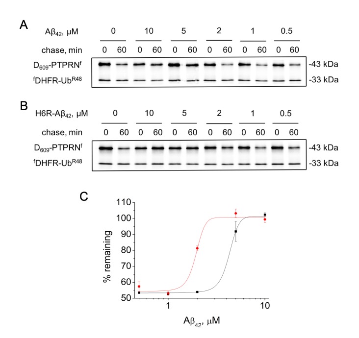
Alzheimer's disease (AD) is accompanied by the dysfunction of intracellular protein homeostasis systems, in particular the ubiquitin-proteasome system (UPS). Beta-amyloid peptide (Aβ), which is involved in the processes of neurodegeneration in AD, is a substrate of this system, however its effect on UPS activity is still poorly explored. Here we found that Aβ peptides inhibited the proteolytic activity of the antiapoptotic Arg/N-end rule pathway that is a part of UPS. We identified arginyltransferase Ate1 as a specific component of the Arg/N-end rule pathway targeted by Aβs. Aβ bearing the familial English H6R mutation, known to cause early-onset AD, had an even greater inhibitory effect on protein degradation through the Arg/N-end rule pathway than intact Aβ. This effect was associated with a significant decrease in Ate1-1 and Ate1-3 catalytic activity. We also found that the loss of Ate1 in neuroblastoma Neuro-2a cells eliminated the apoptosis-inducing effects of Aβ peptides. Together, our results show that the apoptotic effect of Aβ peptides is linked to their impairment of Ate1 catalytic activity leading to suppression of the Arg/N-end rule pathway proteolytic activity and ultimately cell death.

Keywords: Alzheimer’s disease; Ate1; age-related disease; apoptosis; protein degradation.


Conflict of interest statement

CONFLICTS OF INTEREST: The authors declare no conflicts of interests.

Figures

Figure 1
Figure 1
Inhibition effect of Aβ42 (10 μM) on the proteolytic activity of the Arg/N-end rule pathway. (A) Diagram of the fDHFR-UbR48-X609-PTPRNf (X = Asp, Arg-Asp) fusion. Co-translational cleavage of the fusion by deubiquitylases produces a test protein X609-PTPRNf and a stable ’reference’ protein fDHFR-UbR48 at the initially equimolar ratio. (B) Degradation of Asp609-PTPRNf in reticulocyte lysate in the presence or absence of Aβ42. Asp609-PTPRNf was expressed in reticulocyte lysate and co-translationally labeled with 35S-Met for 30 min at 30°C in the presence or absence of Aβ42, followed by a chase, immunoprecipitation with anti-flag M2 antibody, SDS-PAGE, and autoradiography. (C) Same as (B) but with Arg-Asp609-PTPRNf fragment. (D) Quantification of (B). The level of Asp609-PTPRNf was normalized on the level of fDHFR-UbR48. The level of Asp609-PTPRNf detected immediately after stopping of protein expression in reticulocyte lysate (0 min chase) was taken as 100%. "% remaining" is the level of non-degraded Asp609-PTPRNf at shown time points after stopping of protein expression. The absence of Aβ42 - dark-gray column; the presence of Aβ42 – light-gray column. (E) Quantification of (C). Each value is the mean ± SD of at least three independent experiments; *p < 0.01, **p < 0.001.
Figure 2
Figure 2
Effect of different concentrations of amyloid peptides on the proteolytic activity of the Arg/N-end rule pathway. Degradation of 35S-methionine labeled Asp609-PTPRNf in the presence of Aβ42 (A) or H6R-Aβ42 (B) was assayed as described in the legend to Figure 1B. (C) – quantification of (A) and (B) at 60 min chase (Aβ42 – black squares, H6R-Aβ42 – red circles). Each value is the mean expressed as a percentage of the level of Asp609-PTPRNf at 0 min chase ± SD of at least three independent experiments.
Figure 3
Figure 3
Interaction of Aβ42, tRNA, and Ate1. Schematic representation of ELISA assay with immobilized anti-Aβ1-17 antibodies (A) or anti-Aβ36-42 antibodies (B). (C) Detection of tRNA and Ate1 ability to interact with C-terminus of Aβ. (D) Same as (C) but with N-terminal region of Aβ. OD450 – optical density measured at 450 nm. Each value is the mean ± SD of at least four independent experiments; *p < 0.04, **p < 0.01, ***p < 0.001.
Figure 4
Figure 4
Apoptotic effects of Aβ peptides (10 µM, 20 h) on Neuro-2a (A) cells and Ate1 knockout Neuro-2a cells (B). The Annexin-V positive and PI negative cells were considered early apoptotic. Each value is expressed as a percentage of the total number of cells ± SD. The experiments were performed three times in triplicates; *p < 0.01, **p < 0.001.

References

    1. Walsh DM, Selkoe DJ. A critical appraisal of the pathogenic protein spread hypothesis of neurodegeneration. Nat Rev Neurosci. 2016; 17:251–60. 10.1038/nrn.2016.13 - DOI - PMC - PubMed
    1. Khachaturian ZS. Diagnosis of Alzheimer’s disease. Arch Neurol. 1985; 42:1097–105. 10.1001/archneur.1985.04060100083029 - DOI - PubMed
    1. Mirra SS, Hart MN, Terry RD. Making the diagnosis of Alzheimer’s disease. A primer for practicing pathologists. Arch Pathol Lab Med. 1993; 117:132–44. - PubMed
    1. Ji XR, Cheng KC, Chen YR, Lin TY, Cheung CH, Wu CL, Chiang HC. Dysfunction of different cellular degradation pathways contributes to specific β-amyloid42-induced pathologies. FASEB J. 2018; 32:1375–87. 10.1096/fj.201700199RR - DOI - PubMed
    1. Ntsapi C, Lumkwana D, Swart C, du Toit A, Loos B. New Insights Into Autophagy Dysfunction Related to Amyloid Beta Toxicity and Neuropathology in Alzheimer’s Disease. 1st ed. International Review of Cell and Molecular Biology. Elsevier Inc.; 2017. 321–361 p. - PubMed

Publication types

LinkOut - more resources