Review
. 2019 Oct;28(10):2266-2274.
doi: 10.1007/s00586-019-06119-6. Epub 2019 Aug 24.

Measuring and reporting of vertebral endplate bone marrow lesions as seen on MRI (Modic changes): recommendations from the ISSLS Degenerative Spinal Phenotypes Group

Affiliations
Review

Measuring and reporting of vertebral endplate bone marrow lesions as seen on MRI (Modic changes): recommendations from the ISSLS Degenerative Spinal Phenotypes Group

Aaron J Fields et al. Eur Spine J. 2019 Oct.

Abstract

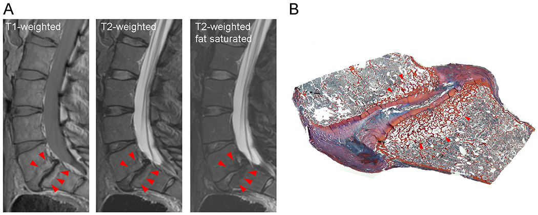
Purpose: The positive association between low back pain and MRI evidence of vertebral endplate bone marrow lesions, often called Modic changes (MC), offers the exciting prospect of diagnosing a specific phenotype of chronic low back pain (LBP). However, imprecision in the reporting of MC has introduced substantial challenges, as variations in both imaging equipment and scanning parameters can impact conspicuity of MC. This review discusses key methodological factors that impact MC classification and recommends guidelines for more consistent MC reporting that will allow for better integration of research into this LBP phenotype.

Methods: Non-systematic literature review.

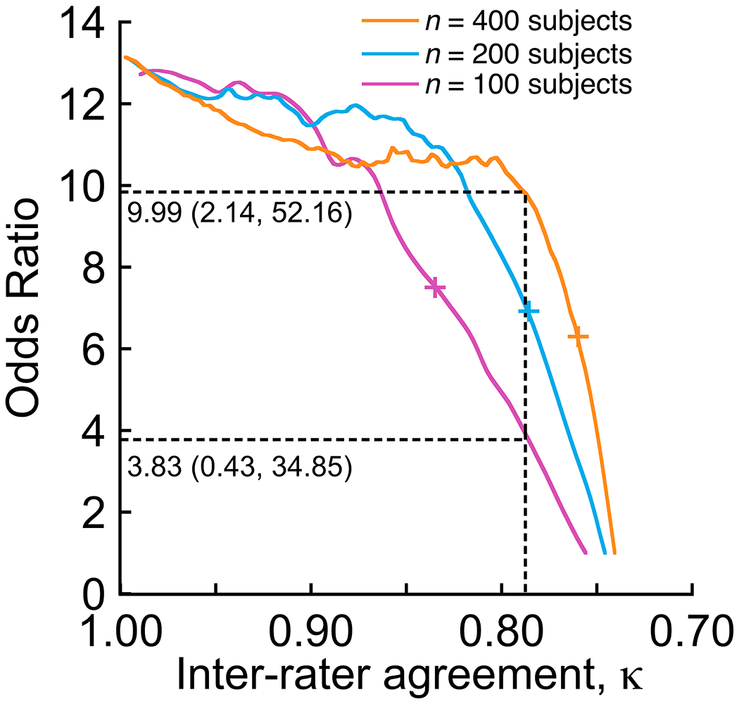
Results: The high diagnostic specificity of MC classification for a painful level contributes to the significant association observed between MC and LBP, whereas low and variable sensitivity underlies the between- and within-study variability in observed associations. Poor sensitivity may be owing to the presence of other pain generators, to the limited MRI resolution, and to the imperfect reliability of MC classification, which lowers diagnostic sensitivity and thus influences the association between MC and LBP. Importantly, magnetic field strength and pulse sequence parameters also impact detection of MC. Advances in pulse sequences may improve reliability and prove valuable for quantifying lesion severity.

Conclusions: Comparison of MC data between studies can be problematic. Various methodological factors impact detection and classification of MC, and the lack of reporting guidelines hinders interpretation and comparison of findings. Thus, it is critical to adopt imaging and reporting standards that codify acceptable methodological criteria. These slides can be retrieved under Electronic Supplementary Material.

Keywords: Bone marrow lesion; Endplate damage; Low back pain; Magnetic resonance imaging; Modic changes.


Figures

Figure 1.
Figure 1.
Hypothetical scenarios show how the reliability of Modic classification, or inter-rater agreement, impacts the relationship between Modic changes and LBP. In each scenario, the actual prevalence of Modic changes in LBP patients (46% [5]) and asymptomatic controls (6% [5]) was simulated, and increasing numbers of false negatives were randomly assigned to the groups (group size = n/2). After each false negative assignment, the inter-rater agreement (κ) and odds ratio was calculated. This simulation was repeated 100 times in order to consider cases where the false negative assignments randomly affected members of each group. The mean odds ratio was calculated for 100 repeat simulations. Plus signs (+) for each scenario indicate the cutoff value of κ that produced a significant odds ratio for each scenario: 100 subjects, κ = 0.837, OR = 7.69 (CI 1.09–56.81); 200 subjects, κ = 0.786, OR = 7.04 (CI 1.03–51.03); 400 subjects, κ = 0.761, OR = 7.27 (CI 1.04–53.53). For κ = 0.788 (vertical dashed line), which was the mean reliability of Modic classification from the six studies summarized in Table 2, the association between Modic changes and LBP ranged from a mean OR = 3.83 (CI 0.43–34.85, not significant) to a mean OR = 9.99 (CI 2.14–52.16, p < 0.001).
Figure 2.
Figure 2.
(A) Type 2 Modic changes seen on sagittal T1- and T2-weighted images of a fresh cadaveric lumbar spine. Use of fat suppression reverses the hyperintense signal on the T2-weighted images. Images were acquired at 3.0T with a fast spin-echo sagittal T2 sequence (repetition time msec/echo time msec 4282/85; 27-cm field of view; 3-mm slice thickness) and a sagittal T1 sequence (556/14, 27-cm field of view, 3-mm slice thickness). (B) Matching sagittal histologic section of L5-S1 level indicating endplate bone marrow lesion with fatty replacement of the hematopoietic elements. Heidenhain tri-chrome stain.
Figure 3.
Figure 3.
(A) Type 1 Modic change contoured on sagittal T1- and fat saturated T2-weighted images from a 52-year-old male subject presenting with chronic LBP. Contouring enables measurement of the size and relative intensity of the Modic change. Images were acquired with a fast spin-echo sagittal T2 sequence (repetition time msec/echo time msec 4933/66; 26-cm field of view; 4-mm slice thickness) and a sagittal T1 sequence (694/15, 26-cm field of view, 4-mm slice thickness). (B) Matching sagittal UTE image showing disruptions in the continuity of the cartilage endplate adjacent to the Modic change. In addition to providing an assessment of cartilage endplate integrity, UTE shows good marrow contrast. Images were acquired with a UTE sequence that combines a nonselective hard pulse and 3D radial acquisition (10/0.236, 19-degree flip angle, 15-cm field of view, 1.5-mm slice thickness). (C) Matching sagittal fat fraction map from water-fat MRI showing reduced fat fraction in the region corresponding to the contour in (A). Compared to site-matched normal regions in the adjacent, non-affected vertebrae, regions with type 1 Modic changes had lower fat fraction, suggesting that water-fat MRI is sensitive to the fibrovascular replacement of the normal bone marrow that occurs with this type of endplate bone marrow lesion. Data are shown for three subjects presenting with chronic LBP, and fat fractions are shown as mean ± SD for three ROIs (3-mm diameter) per region. Images were acquired with a 3D spoiled gradient recalled sequence (10/1.3, 3-degree flip angle, 22-cm field of view, 4-mm slice thickness) with six echoes and iterative decomposition of water and fat with echo asymmetry and least squares estimation (IDEAL). All images were acquired at 3.0T.

References

    1. Modic MT, Steinberg PM, Ross JS, Masaryk TJ, Carter JR (1988) Degenerative disk disease: assessment of changes in vertebral body marrow with MR imaging. Radiology 166 (1 Pt 1):193–199 - PubMed
    1. de Roos A, Kressel H, Spritzer C, Dalinka M (1987) MR imaging of marrow changes adjacent to end plates in degenerative lumbar disk disease. AJR Am J Roentgenol 149 (3):531–534. doi:10.2214/ajr.149.3.531 - DOI - PubMed
    1. Modic MT, Masaryk TJ, Ross JS, Carter JR (1988) Imaging of degenerative disk disease. Radiology 168 (1):177–186. doi:10.1148/radiology.168.1.3289089 - DOI - PubMed
    1. Jensen RK, Leboeuf-Yde C (2011) Is the presence of modic changes associated with the outcomes of different treatments? A systematic critical review. BMC Musculoskelet Disord 12:183. doi:10.1186/1471-2474-12-183 - DOI - PMC - PubMed
    1. Jensen TS, Karppinen J, Sorensen JS, Niinimaki J, Leboeuf-Yde C (2008) Vertebral endplate signal changes (Modic change): a systematic literature review of prevalence and association with non-specific low back pain. Eur Spine J 17 (11):1407–1422. doi:10.1007/s00586-008-0770-2 - DOI - PMC - PubMed

Publication types

MeSH terms