. 2020 Jun;11(3):365-376.
doi: 10.1007/s12975-019-00725-8. Epub 2019 Aug 24.

A Brain-Targeted Orally Available ROCK2 Inhibitor Benefits Mild and Aggressive Cavernous Angioma Disease

Affiliations

A Brain-Targeted Orally Available ROCK2 Inhibitor Benefits Mild and Aggressive Cavernous Angioma Disease

Lisa McKerracher et al. Transl Stroke Res. 2020 Jun.

Abstract

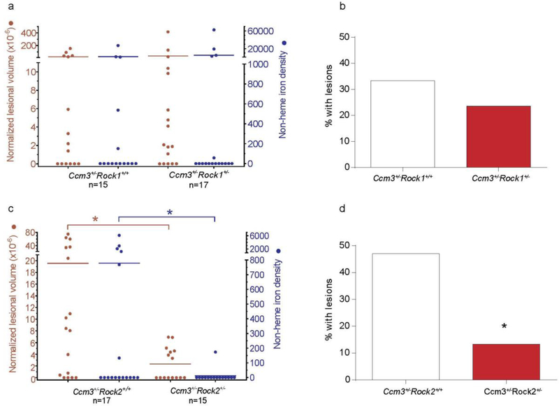
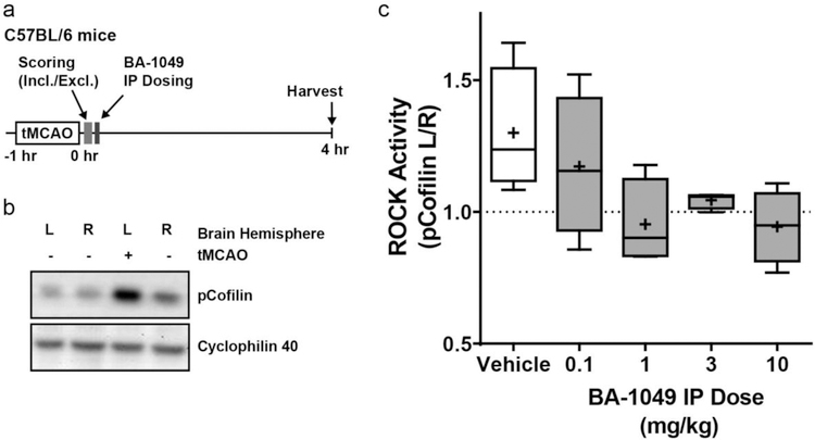
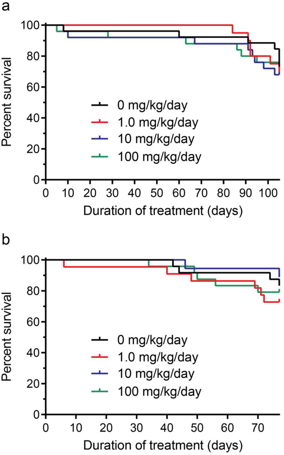
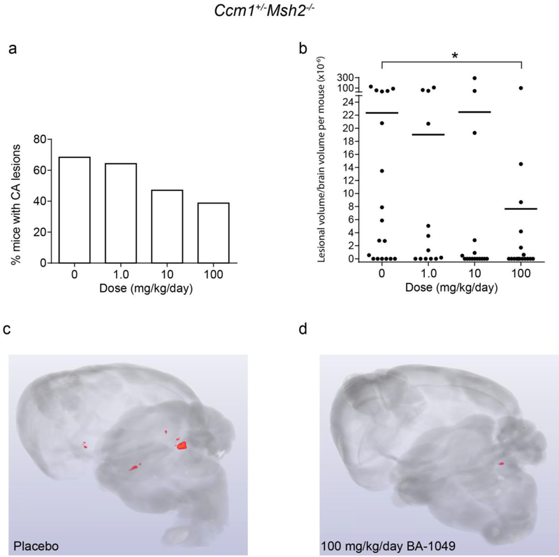
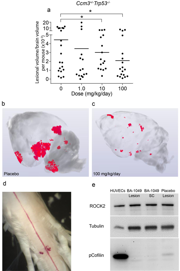
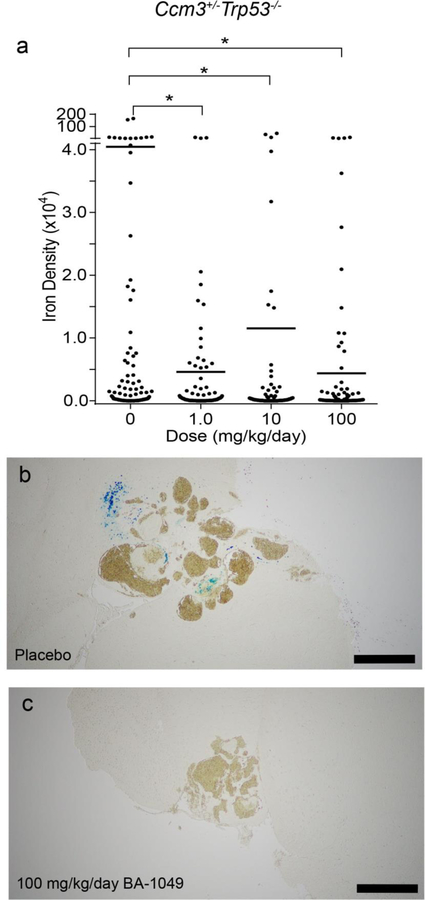
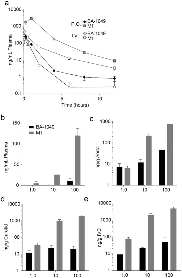
Cavernous angioma (CA) is a vascular pathology caused by loss of function in one of the 3 CA genes (CCM1, CCM2, and CCM3) that result in rho kinase (ROCK) activation. We investigated a novel ROCK2 selective inhibitor for the ability to reduce brain lesion formation, growth, and maturation. We used genetic methods to explore the use of a ROCK2-selective kinase inhibitor to reduce growth and hemorrhage of CAs. The role of ROCK2 in CA was investigated by crossing Rock1 or Rock2 hemizygous mice with Ccm1 or Ccm3 hemizygous mice, and we found reduced lesions in the Rock2 hemizygous mice. A ROCK2-selective inhibitor, BA-1049 was used to investigate efficacy in reducing CA lesions after oral administration to Ccm1+/- and Ccm3+/- mice that were bred into a mutator background. After assessing the dose range effective to target brain endothelial cells in an ischemic brain model, Ccm1+/- and Ccm3+/- transgenic mice were treated for 3 (Ccm3+/-) or 4 months (Ccm1+/-), concurrently, randomized to receive one of three doses of BA-1049 in drinking water, or placebo. Lesion volumes were assessed by micro-computed tomography. BA-1049 reduced activation of ROCK2 in Ccm3+/-Trp53-/- lesions. Ccm1+/-Msh2-/- (n=68) and Ccm3+/-Trp53-/- (n=71) mice treated with BA-1049 or placebo showed a significant dose-dependent reduction in lesion volume after treatment with BA-1049, and a reduction in hemorrhage (iron deposition) near lesions at all doses. These translational studies show that BA-1049 is a promising therapeutic agent for the treatment of CA, a disease with no current treatment except surgical removal of the brain lesions.

Keywords: Cerebral cavernous malformation; Hemangioma; Rho kinase; Therapeutics; Vascular permeability.


Conflict of interest statement

Competing interests

Dr. McKerracher is CEO of BioAxone, holds an ownership interest in the company and has a significant competing interest. Drs. Matthew Abbinanti, Ken Rosen and Joerg Ruschel were employees of BioAxone and have a modest conflict of interest. Drs. Doug Marchuk and Issam Awad were recipients of sponsored research through the SBIR grant to BioAxone. The other authors declare no conflict of interest.

Figures

Fig. 1
Fig. 1
The effect of Rock haploinsufficiency on cavernous angioma lesion burden and hemorrhage in Ccm3+/− models. Rock1 haploinsufficiency did not affect (a) lesion burden (left axis, red) and non-heme iron deposition (right axis, blue) per mouse, and (b) the percentage of mice with mature, multi-cavernous Stage 2 lesions. In contrast, Rock2 haploinsufficiency significantly decreased (c) lesion burden (P=0.011) and non-heme iron deposition (P=0.049) per mouse, and (d) the percentage of mice with Stage 2 lesions (P=0.040). The Mann Whitney U test (a, c) and χ2 test (b, d) were used to assess for significant differences. *P<0.05.
Fig. 2
Fig. 2
Dose-response for BA-1049 to reduces ROCK hyperactivation in mice. (a) Experimental design of 60 minute transient middle cerebral artery occlusion (tMCAO) in C57BL/6 mice used to examine dose-response after a single application of BA-1049. (b) Increase in phosphorylated Cofilin (pCofilin) is detected in the left (L) ischemic side in brain after tMCAO compared to the right (R) contralateral side. (c) Dose-response determined as ratio of pCofilin expression on the ischemic left side of brain compared to right brain (pCofilin L/R) after MCAO lesion and treated with vehicle, 0.1, 1, 3 or 10 mg/kg BA-1049 HCl. There was a trend towards an effect of BA-1049 on ROCK activation (χ2(4) = 8.757, P=0.0675, n=4 animals per group).
Fig. 3
Fig. 3
Attrition in cavernous angioma models treated with BA-1049. (a) Ccm1+/−Msh2−/− models were treated with 0 (n=26), 1.0 (n=20), 10 (n=25) or 100 (n=25) mg/kg/day BA-1049 from weaning to the earliest age for the end of treatment (105 days of age). (b) Ccm3+/−Trp53−/− models were treated with 0 (n=24), 1.0 (n=22), 10 (n=18) or 100 (n=24) mg/kg/day BA-1049 from weaning to the earliest age for the end of treatment (77 days of age). Kaplan-Meier plots show no significant effect of treatment on survival compared with placebos. The log-rank (Mantel-Cox) test was used to assess for significant differences.
Fig. 4
Fig. 4
The effect of BA-1049 therapy on cavernous angioma (CA) lesion burden in Ccm1+/−Msh2−/− models. (a) There was a trend (P=0.063) for fewer mice harboring CA lesions that were treated 100 mg/kg/day BA-1049 compared with placebos. (b) Treatment with 100 mg/kg/day BA-1049 significantly decreased lesion burden compared with placebos (P=0.022). Representative micro-computed tomography show (c) more prevalent CA lesions (red areas) in placebos than in (d) mice treated with 100 mg/kg/day BA-1049. No significant effect was found with treatment at lower doses of BA-1049. The χ2 (a) test and Mann Whitney U test (b) were used to assess for significance. *P<0.05. There were 18, 13, 16 and 17 animals per group treated with 0, 1.0, 10 and 100 mg/kg/day BA-1049 respectively.
Fig. 5
Fig. 5
The effect of BA-1049 therapy on the Ccm3+/−Trp53−/− model. (a) BA-1049 therapy at 10 and 100 mg/kg/day significantly decreased cavernous angioma (CA) lesion burden (P=0.002, P=0.003 vs. placebo, respectively). There were 20, 15, 16 and 18 animals per group treated with 0, 1.0, 10 and 100 mg/kg/day BA-1049 respectively. Representative micro-computed tomography show (b) more prevalent CA lesions (red areas) in placebos than in (c) mice treated with 100 mg/kg/day BA-1049. (d) A spinal cord (SC) from a Ccm3+/−Trp53−/− mouse showing CA lesion on the ventral surface. (e) Western blot probed with antibodies to ROCK2, tubulin as control and phosphorylated Cofilin (pCofilin) as a biomarker of ROCK activation. Human umbilical vein ECs (HUVECs) treated with lipoprotein(a) to activate ROCK2 were used as a ROCK activation control. Lesion and SC samples from a mouse treated with 100 mg/kg/day BA-1049 is shown and compared with a mouse given placebo. The 2-sided Conover 2-sample test was used to assess for significant differences. *P<0.05.
Fig. 6
Fig. 6
The effect of BA-1049 therapy on hemorrhage in the Ccm3+/−Trp53−/− model. (a) BA-1049 therapy at 1.0, 10 and 100 mg/kg/day significantly decreased non-heme iron per Stage 2 lesional area compared with placebos (P=0.037, P=0.0015, P=0.00003 vs. placebo, respectively). There were 108, 85, 95 and 112 mature, multicavernous Stage 2 lesions per group in animals treated with 0, 1.0, 10 and 100 mg/kg/day BA-1049 respectively. Representative cavernous angioma lesions show (b) the presence of non-heme iron (blue Perls stain) in placebos and (c) non-heme iron lacking in mice treated with 100 mg/kg/day. The Mann Whitney U test was used to assess for significant differences. Bar, 500 µm. *P<0.05.
Fig. 7
Fig. 7
Pharmacokinetics and exposure of BA-1049 and M1 in Ccm mice. (a) Pharmacokinetic analysis of BA-1049 (circles) and metabolite M1 (squares) after oral (P.O.; 30 mg/kg; n = 4 at each time point) and intravenous (I.V.; 5 mg/kg; n = 4) delivery of BA-1049 in mice. (b) BA-1049 (gray) and M1 (black) content in plasma of Ccm1 and Ccm3 mice after 4 or 3 months of treatment (respectively) of 1.0 (n = 11), 10 (n = 14), or 100 (n = 38) mg/kg/day BA-1049 in drinking water. (c) BA-1049 (gray) and M1 (black) content in aorta of Ccm1 and Ccm3 mice after 4 or 3 months of treatment (respectively) of 1.0 (n = 9), 10 (n = 11), or 100 (n = 35) mg/kg/day BA-1049 in drinking water. (d) BA-1049 (gray) and M1 (black) content in carotid of Ccm1 and Ccm3 mice after 4 or 3 months of treatment (respectively) of 1.0 (n = 8), 10 (n = 10), or 100 (n = 25) mg/kg/day BA-1049 in drinking water. (e) BA-1049 (gray) and M1 (black) content in inferior vena cava (IVC) of Ccm1 and Ccm3 mice after 4 or 3 months of treatment (respectively) of 1.0 (n = 8), 10 (n = 20), or 100 (n = 23) mg/kg/day BA-1049 in drinking water.

References

    1. Al-Shahi Salman R, Hall JM, Horne MA, Moultrie F, Josephson CB, Bhattacharya JJ et al. Untreated clinical course of cerebral cavernous malformations: a prospective, population-based cohort study. Lancet Neurol 2012;11(3):217–24. 10.1016/S1474-4422(12)70004-2. - DOI - PMC - PubMed
    1. Riant F, Bergametti F, Ayrignac X, Boulday G, Tournier-Lasserve E. Recent insights into cerebral cavernous malformations: the molecular genetics of CCM. FEBS J 2010;277(5):1070–5. 10.1111/j.1742-4658.2009.07535.x. - DOI - PubMed
    1. Leblanc GG, Golanov E, Awad IA, Young WL, Biology of Vascular Malformations of the Brain NWC. Biology of vascular malformations of the brain. Stroke 2009;40(12):e694–702. 10.1161/STROKEAHA.109.563692. - DOI - PMC - PubMed
    1. Al-Holou WN, O’Lynnger TM, Pandey AS, Gemmete JJ, Thompson BG, Muraszko KM et al. Natural history and imaging prevalence of cavernous malformations in children and young adults. J Neurosurg Pediatr 2012;9(2):198–205. 10.3171/2011.11.PEDS11390. - DOI - PubMed
    1. Plummer NW, Gallione CJ, Srinivasan S, Zawistowski JS, Louis DN, Marchuk DA. Loss of p53 sensitizes mice with a mutation in Ccm1 (KRIT1) to development of cerebral vascular malformations. Am J Pathol 2004;165(5):1509–18. 10.1016/S0002-9440(10)63409-8. - DOI - PMC - PubMed

Publication types

MeSH terms

LinkOut - more resources