. 2019 Sep 5;105(3):493-508.
doi: 10.1016/j.ajhg.2019.07.007. Epub 2019 Aug 22.

Aberrant Function of the C-Terminal Tail of HIST1H1E Accelerates Cellular Senescence and Causes Premature Aging

Affiliations

Aberrant Function of the C-Terminal Tail of HIST1H1E Accelerates Cellular Senescence and Causes Premature Aging

Elisabetta Flex et al. Am J Hum Genet. .

Abstract

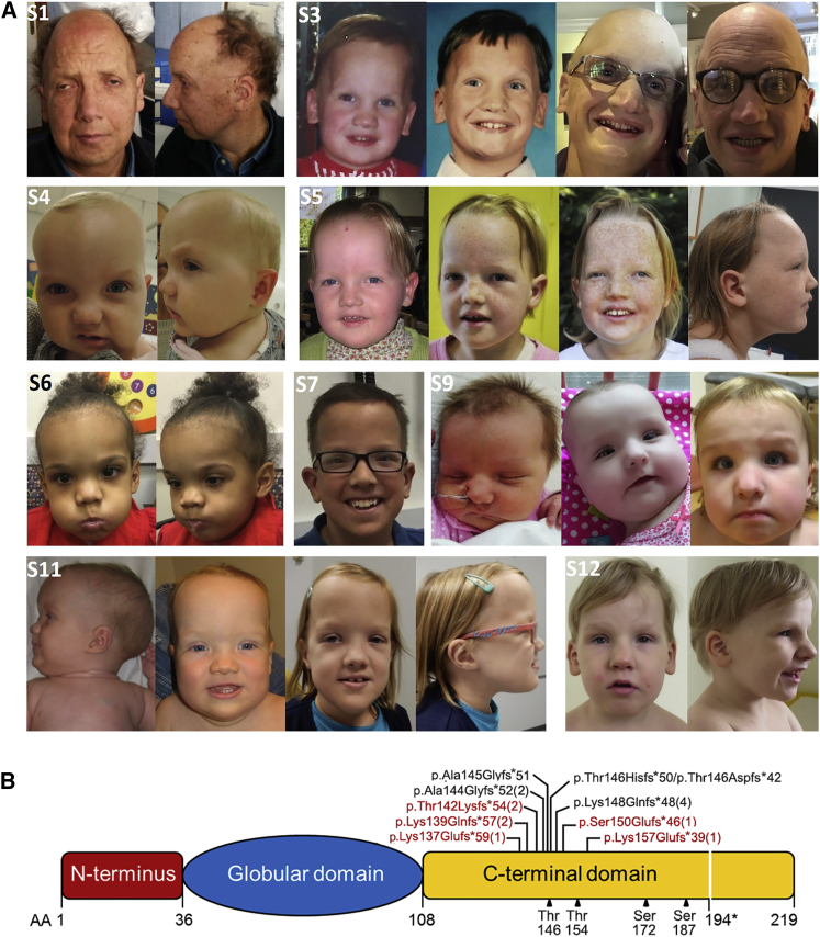
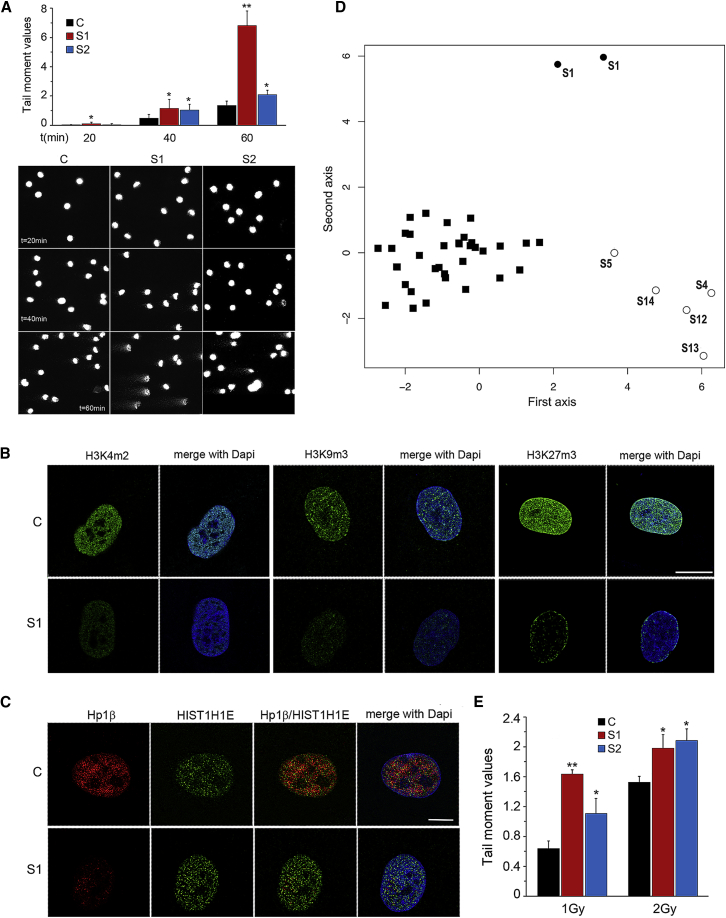
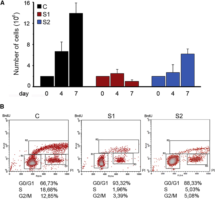
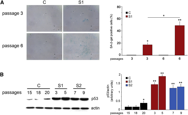
Histones mediate dynamic packaging of nuclear DNA in chromatin, a process that is precisely controlled to guarantee efficient compaction of the genome and proper chromosomal segregation during cell division and to accomplish DNA replication, transcription, and repair. Due to the important structural and regulatory roles played by histones, it is not surprising that histone functional dysregulation or aberrant levels of histones can have severe consequences for multiple cellular processes and ultimately might affect development or contribute to cell transformation. Recently, germline frameshift mutations involving the C-terminal tail of HIST1H1E, which is a widely expressed member of the linker histone family and facilitates higher-order chromatin folding, have been causally linked to an as-yet poorly defined syndrome that includes intellectual disability. We report that these mutations result in stable proteins that reside in the nucleus, bind to chromatin, disrupt proper compaction of DNA, and are associated with a specific methylation pattern. Cells expressing these mutant proteins have a dramatically reduced proliferation rate and competence, hardly enter into the S phase, and undergo accelerated senescence. Remarkably, clinical assessment of a relatively large cohort of subjects sharing these mutations revealed a premature aging phenotype as a previously unrecognized feature of the disorder. Our findings identify a direct link between aberrant chromatin remodeling, cellular senescence, and accelerated aging.

Keywords: HIST1H1E; accelerated aging; cellular senescence; chromatin compaction; chromatin dynamics; chromatin remodeling; linker histone; linker histone H1.4; methylation profiling; replicative senescence.


Conflict of interest statement

A.Be. and R.P. declare no additional conflicts of interest beyond their employment affiliation. All of the other authors declare no competing interests.

Figures

Figure 1
Figure 1
Facial Appearance of Subjects with HIST1H1E Frameshift Mutations and Protein Structure (A) In affected individuals, facial appearance is characterized by a high anterior hairline, prominent forehead, bitemporal narrowing, sparse temporal hair, hypertelorism, hooded eyelids, short palpebral fissures, a high and broad nasal bridge, and a full nasal tip; small, widely spaced teeth; and low-set ears. A facial appearance compatible with a more advanced age (e.g., hypotrichosis and ptosis) is evident in S1 and S3. (S1, 49 years; S3, 30 years at last evaluation; S4, 14 months; S5, 12 years at last evaluation; S6, 3 years; S7, 12 years; S9, 2 years at last evaluation; S11, 6 years at last evaluation; and S12, 4 years) (B) Schematic diagram representing the HIST1H1E structure, which is composed of a globular domain flanked by N- and C-terminal tails. The position of the disease-causing frameshift mutations is shown above the cartoon. (Novel mutations are highlighted in red.) The number of independent cases identified in the present study is in brackets. The domain boundaries and cyclin-dependent kinase phosphorylation sites (black triangles) are reported below the cartoon. All mutations are expected to result in a shorter protein with an identical divergent C-terminal tail. (The new stop codon is shown below the cartoon, 194.)
Figure 2
Figure 2
SCGE Assay, Immuno-Fluorescence Studies, and Methylation Profiling Analysis (A) Increasing electrophoresis run times (20, 40, and 60 min) highlighted significant differences in relaxation of DNA supercoiling between fibroblasts from control and from affected individuals. DNA migration was quantified as Tail moment values, which are defined as the products of the tail length and the fraction of total DNA in the tail (upper panel). Nucleoids of cells from subjects S1 and S2 showed a significantly higher Tail moment value (p < 0.05, ∗∗p < 0.01; two-tailed Student’s t test). For each experimental point, at least 75 cells were analyzed. Values are mean ± SEM of three independent experiments. Representative images of nucleoids from fibroblasts from control and affected individuals at each run time are shown (lower panel). (B) Confocal laser scanning microscopy (CLSM) observations document overall decreased amounts of H3K4me2, H3K9me3, and H3K27me3 staining (green) in S1 cells compared to control cells. Nuclei were stained with DAPI. Images are representative of >200 analyzed cells. Scale bars represent to 8 μm. (C) Fibroblasts from subject S1 show a decreased amount of HP1β compared to control cells (WT). Cells were stained with antibodies against HP1β (red) and HIST1H1E (H1.4) (green); DNA are DAPI-stained (blue). Scale bars represent 7 μm. (D) Multidimensional scaling plot of genome-wide methylation analysis using the top 1,000 most variable probes among samples. The plot shows the distinct methylation profiles of pediatric (open circles) and adult (S1, duplicate) (filled circles) affected individuals, compared to healthy controls (filled squares). (E) DNA damage was induced by 1 or 2 Gy γ-ray irradiation. Tail moment values indicate the amount of radiation-induced DNA damage measured by SCGE assay immediately after treatment. S1 and S2 fibroblasts showed a higher sensitivity to γ-ray irradiation (p < 0.02, ∗∗p < 0.001; two-tailed Student’s t test). For each experimental point, at least 75 cells were analyzed. Values are means ± SEM of three independent experiments.
Figure 3
Figure 3
Proliferation Assay and Cell-Cycle Progression (A) Cells were seeded at 200,000 cells/well in a six-well plate and incubated a 37°C. Cell numbers (mean of three replicates ± SD) were counted by trypan blue exclusion assay after four and seven days. A significantly decreased proliferation rate was observed in the fibroblast lines from the two unrelated affected individuals (S1 and S2). Cells from subject S1 show a permanent cell growth arrest. (B) Cell cycle phases of S1/S2’s (right) and control (left) fibroblasts as measured by BrdU incorporation and propidium iodide (PI) flow cytometry analysis. The upper box identifies cells incorporating BrdU (S phase), the lower left box identifies G0/G1 cells and the lower right box represents G2/M cells. One of three independent experiments is reported with the percentage of cells in each cell cycle phase.
Figure 4
Figure 4
Defective HISTH1E Function Results in Altered SA-β-gal Activity and p53 Expression Level (A) Representative images (left) and quantification (right) of SA-β-gal activity evaluated on S1 and control (C) fibroblasts at different culture passages. The significance was measured by one-way Anova with Tukey’s multiple comparison test (p < 0.01, ∗∗p < 0.0001). (B) Compared to control cell lysates, fibroblast lysates from individuals S1 and S2 showed enhanced TP53 protein levels at earlier passages. Representative blots (left) and mean ± SD densitometry values (right) of three independent experiments are shown (p < 0.05, ∗∗p < 0.002; two-tailed Student’s t test).
Figure 5
Figure 5
Defective HISTH1E Function Results in Aberrant Nuclear Morphology That Is Exacerbated over Cell-Culture Passages (A) CLSM analysis was performed in steady-state (left) and synchronized (right) skin fibroblasts induced to divide after being treated with thymidine/nocodazole and recovered with fresh medium. The panels show an aberrant nuclear morphology in cells from subject S1. Whereas control cells proceed through mitosis (representative metaphases are shown), S1 fibroblasts fail to progress. Experiments were carried out at early passages (passage 3). Cells were stained with an antibody against lamin A/C (red) and DAPI (blue). Images are representative of >200 analyzed cells. Scale bars represent 7 μm. (B) CLSM analysis was performed on S1 and control fibroblasts seeded at different culture passages. The panels show an aberrant nuclear morphology at early passages in subject S1’s cells compared to control cells, which were seeded at late passages (passage 16). Percentages refer to the number of cells with aberrant nuclear morphology. Cells were stained as above. Images are representative of >200 analyzed cells. Scale bars represent 27 μm.
Figure 6
Figure 6
Defective HIST1H1E Function Results in Nucleolar Fragmentation and Increased 18S and 28S rRNA Levels (A) CLSM observations were performed on S1 and control (C) fibroblasts. Panels show a significant decrease in C3 clone antibody staining in S1 fibroblasts, revealing nucleolar fragmentation. Nuclei were stained with DAPI. Images are representative of >200 analyzed cells. Scale bars represent 9 μm. (B) Total RNA was extracted from the same amount of S1 and C cells at different cellular passages. Three μl of total RNA was loaded for size separation on 1% agarose gel and stained with ethidium bromide. Increased amounts (compared to those in control cells) of both 28S and 18S rRNA codifying for ribosomal subunits are evident in fibroblasts from subject S1.

Similar articles

Cited by

References

    1. Wolffe A. Academic Press; London: 1998. Chromatin: Structure and function.
    1. Luger K., Mäder A.W., Richmond R.K., Sargent D.F., Richmond T.J. Crystal structure of the nucleosome core particle at 2.8 A resolution. Nature. 1997;389:251–260. - PubMed
    1. Harshman S.W., Young N.L., Parthun M.R., Freitas M.A. H1 histones: current perspectives and challenges. Nucleic Acids Res. 2013;41:9593–9609. - PMC - PubMed
    1. Hergeth S.P., Schneider R. The H1 linker histones: multifunctional proteins beyond the nucleosomal core particle. EMBO Rep. 2015;16:1439–1453. - PMC - PubMed
    1. Crane-Robinson C. Linker histones: History and current perspectives. Biochim. Biophys. Acta. 2016;1859:431–435. - PubMed

Publication types