. 2019 Aug 25;11(16):5924-5942.
doi: 10.18632/aging.102150. Epub 2019 Aug 25.

Cognitive impairment in heart failure is associated with altered Wnt signaling in the hippocampus

Affiliations

Cognitive impairment in heart failure is associated with altered Wnt signaling in the hippocampus

Camilo Toledo et al. Aging (Albany NY). .

Abstract

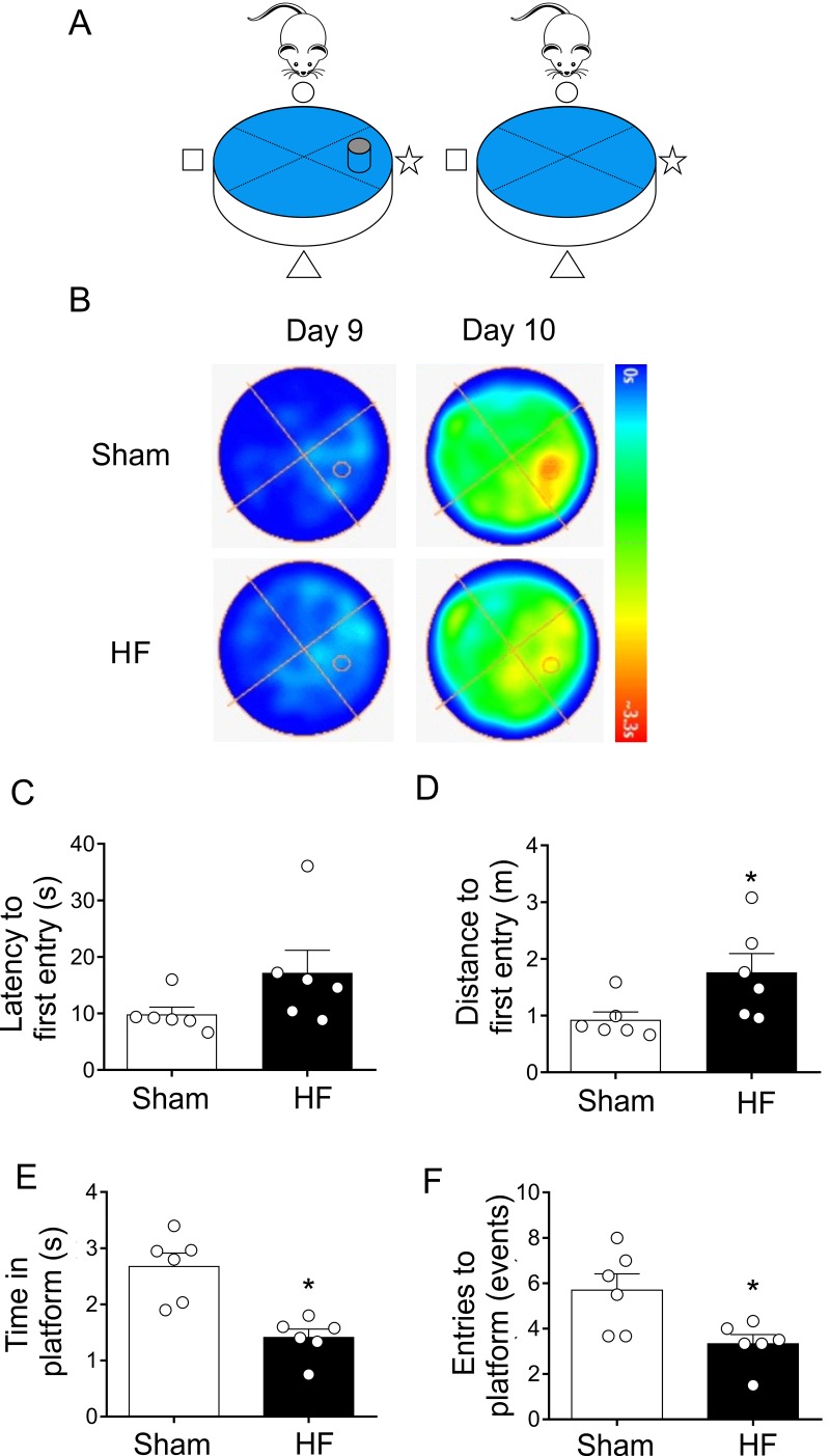
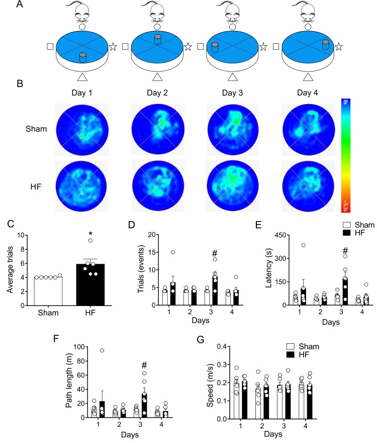
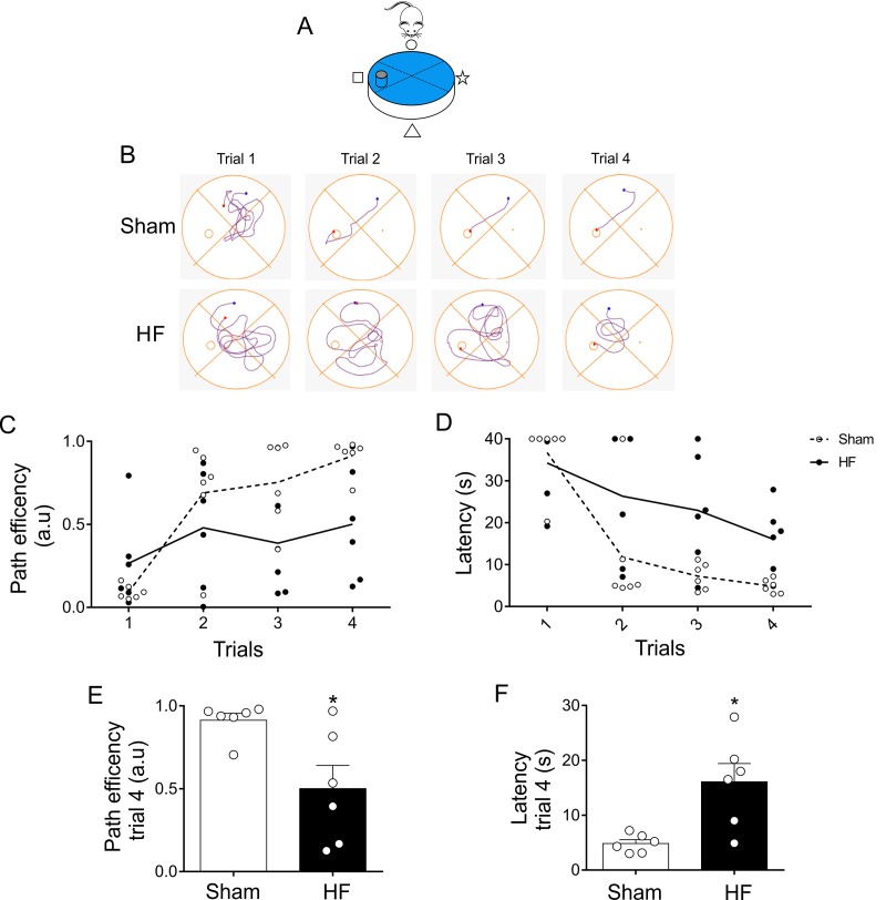
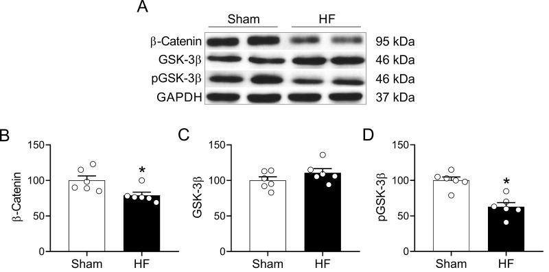
Age represents the highest risk factor for death due to cardiovascular disease. Heart failure (HF) is the most common cardiovascular disease in elder population and it is associated with cognitive impairment (CI), diminishing learning and memory process affecting life quality and mortality in these patients. In HF, CI has been associated with inadequate O2 supply to the brain; however, an important subset of HF patients displays CI with almost no alteration in cerebral blood flow. Importantly, nothing is known about the pathophysiological mechanisms underpinning CI in HF with no change in brain tissue perfusion. Here, we aimed to study memory performance and learning function in a rodent model of HF that shows no change in blood flow going to the brain. We found that HF rats presented learning impairments and memory loss. In addition, HF rats displayed a decreased level of Wnt/β-catenin signaling downstream elements in the hippocampus, one pathway implicated largely in aging diseases. Taken together, our results suggest that in HF rats CI is associated with dysfunction of the Wnt/β-catenin signaling pathway. The mechanisms involved in the alterations of Wnt/β-catenin signaling in HF and its contribution to the development/maintenance of CI deserves future investigations.

Keywords: Wnt signaling pathway; aging; cognitive impairment; heart failure.


Conflict of interest statement

CONFLICTS OF INTEREST: Authors have no conflicts of interest to declare.

Figures

Figure 1
Figure 1
Study timeline illustrating experimental design. (A) At week 0, heart failure and Sham surgery were made. After 4 weeks, heart failure rats were evaluated by echocardiography to determine cardiac diameters. At 8-weeks, echocardiography, arterial doppler and procedures for assessing spatial and related forms of learning and memory were performed. (B) To determine spatial acquiring learning, rats were trained for two day, then tested for five consecutive days, followed by two days of rest, and three more days of testing. The 10th day, the platform was removed. (C) Followed spatial acquiring learning and after 2 days of rest, same set of rats performed memory flexibility test for four consecutive days and the position of the platform was changed daily.
Figure 2
Figure 2
Carotid artery blood flow in high output heart failure. (A) Representative image of ultrasound scanning showing carotid artery in one Sham rat and one HF rat (B) Representative traces of carotid artery blood flow, assessed by Doppler at rest in one Sham rat and one HF rat. (C) Summary data showing carotid artery diameter. (D) Summary data showing carotid artery blood flow. Note that blood flow to the brain region was unaltered between groups. Values are means ± S.E.M. *p < 0.05 vs. Sham. Unpaired t-test. n=6 rats per group.
Figure 3
Figure 3
Heart failure (HF) rats display spatial learning decline. (A) Representative cartoon showing the rat and platform position in the pool. (B) Representative trajectory heat maps obtained from one Sham rat and one HF rat during Water Maze test between 1 to 5 days. Pseudocolor intensity indicate the time latency that the rat remained swimming in the pool searching for the platform. It is noteworthy that HF rats spends more time searching the platform across all experimental days compared to Sham animals. (C) Summary data of test duration during all experimental days. Note that HF rats required more time to find the platform. (D) Summary data of mean distance travelled during all experimental days. HF rats travel more distance compared to Sham animals. (E) Compared to Sham rats, HF animals display a decreased path efficiency in day 3 and 5. (F) Sham and HF rats display similar speed in all experimental days. &, p<0.05 vs. Sham day 1; +, p<0.05 vs. HF day 1; #, p<0.05 vs. Sham; two-way ANOVA following Holm Sidak test. n=6 rats per group.
Figure 4
Figure 4
Heart failure (HF) rats display similar reference spatial memory compared to control rats. (A) Representative cartoon showing the rat and platform position in the pool. (B) Representative trajectory heat maps obtained from one Sham rat and one HF rat during Water Maze test between day 5 and day 8. Pseudocolor intensity indicates the time latency that the rat remained swimming in the pool searching for the platform. HF rats spends more time searching the platform in day 5; however, this behavior was absent in day 8. (C) Summary data showing that at day 5 HF rats need more time to find the platform; however, this effect was absent at day 8. (D) Summary data showing mean distance travelled during day 5 and day 8. At day 5 HF rats travel more distance compared to Sham animals. No significant differences in traveled distance was found between groups at day 8. (E, F) Sham and HF rats display similar path efficiency and speed in day 5 and day 8 of the test. #, p<0.05 vs. Sham day 5; two-way ANOVA following Holm Sidak test. n=6 rats per group.
Figure 5
Figure 5
Heart failure (HF) rats showed consolidation memory decline. (A) Representative cartoon showing the rat and platform position in the pool. (B) Representative trajectory heat maps obtained from one Sham rat and one HF rat during Water Maze test during day 9 and 10. Contrary to day 9, in day 10 the platform was removed. Pseudocolor intensity indicate the time latency that the rat remained swimming and searching for the platform. Contrarily to Sham rats, HF animals showed less persistence in the place where the platform was originally located. (C) Summary data showing latency to first entry to the place where the platform was located (day 9). (D) Summary data showing the distance for first entry to the place where the platform was located (day 9). HF rats travelled more distance to first entry compared to Sham animals. (E, F) HF rats display a significant decrease in the time and entries to the place where the platform was located (compared to day 9). Values are means ± S.E.M. *, p<0.05, Unpaired t-test. n=6 rats per group.
Figure 6
Figure 6
Memory flexibility was impaired in heart failure (HF) rats. (A) Representative cartoon showing the rat and different platform positions within the pool. (B) Representative trajectory heat maps obtained from one Sham rat and one HF rat during day 1 and day 4 of memory flexibility test. Note that during this test the platform position changed every day of the test. Pseudocolor intensity indicate the time latency the rat remained swimming in the pool searching for the platform. Contrarily to day 1, 2 and 4, at day 3 HF animals showed less latency in the platform position. (C) Average trials between day 1 and 4. Note that HF rats needed more trials to accomplish the task compared to Sham rats. (D) Summary data showing the number trials in each experimental day. (E, F) Summary data of the sum of latencies and paths length from each trial by day. Note that at day 3, HF rats display an increase test duration and distance travelled compared to Sham animals. (G) Average speed was not different in all experimental days between Sham and HF rats. Values are means ± S.E.M. *, p<0.05, Unpaired t-test; #, p<0.05 vs. Sham; two-way ANOVA following Holm Sidak test. n=6 rats per group.
Figure 7
Figure 7
Memory flexibility at day 3 was compromised in heart failure (HF) rats. (A) Representative cartoon showing the rat and platform position at day 3. (B) Representative trajectory maps obtained from one Sham rat and one HF rat during day 3 of memory flexibility test. (C) Summary data showing average paths efficiency in each trial during day 3 of testing in Sham (white circle) and HF (black circle) rats. (D) Summary data showing average latencies from each trial during day 3 in Sham (white circle) and HF (black circle) rats. (E, F) Summary data of average paths efficiency and latencies at trial four (n=6 per group). Note that HF rats display an increase in the number of trials needed to complete the test, as well as an increase in the duration of each trial. Values are means ± S.E.M. *, p<0.05 vs. Sham, Unpaired t-test. n=6 rats per group.
Figure 8
Figure 8
HF rats display alterations in the Wnt signaling pathway in the hippocampus. (A) Representative immunoblots showing the expression levels of active β-catenin, total GSK-3β and the inhibited form of GSK-3β (phospho-Ser9) in hippocampus micro-punches obtained from Sham rats and HF rats. (B) Summary data showing densitometric analysis of β-catenin, (C) total GSK-3β (D) and p-GSK-3β normalized against housekeeping protein GAPDH. Values are means ± S.E.M. *, p<0.05, Unpaired t-test. n=6 rats per group.

References

    1. Ambrosy AP, Gheorghiade M, Chioncel O, Mentz RJ, Butler J. Global perspectives in hospitalized heart failure: regional and ethnic variation in patient characteristics, management, and outcomes. Curr Heart Fail Rep. 2014; 11:416–27. 10.1007/s11897-014-0221-9 - DOI - PubMed
    1. Go YY, Allen JC, Chia SY, Sim LL, Jaufeerally FR, Yap J, Ching CK, Sim D, Kwok B, Liew R. Predictors of mortality in acute heart failure: interaction between diabetes and impaired left ventricular ejection fraction. Eur J Heart Fail. 2014; 16:1183–89. 10.1002/ejhf.119 - DOI - PubMed
    1. Cacciatore F, Abete P, Ferrara N, Calabrese C, Napoli C, Maggi S, Varricchio M, Rengo F, and Osservatorio Geriatrico Campano Study Group. Congestive heart failure and cognitive impairment in an older population. J Am Geriatr Soc. 1998; 46:1343–48. 10.1111/j.1532-5415.1998.tb05999.x - DOI - PubMed
    1. Cameron J, Gallagher R, Pressler SJ. Detecting and Managing Cognitive Impairment to Improve Engagement in Heart Failure Self-Care. Curr Heart Fail Rep. 2017; 14:13–22. 10.1007/s11897-017-0317-0 - DOI - PubMed
    1. Harkness K, Demers C, Heckman GA, McKelvie RS. Screening for cognitive deficits using the Montreal cognitive assessment tool in outpatients ≥65 years of age with heart failure. Am J Cardiol. 2011; 107:1203–07. 10.1016/j.amjcard.2010.12.021 - DOI - PubMed

Publication types