. 2019 Aug 9:10:876.
doi: 10.3389/fphar.2019.00876. eCollection 2019.

Specific Inhibition of CYP4A Alleviates Myocardial Oxidative Stress and Apoptosis Induced by Advanced Glycation End-Products

Affiliations

Specific Inhibition of CYP4A Alleviates Myocardial Oxidative Stress and Apoptosis Induced by Advanced Glycation End-Products

Rui Wang et al. Front Pharmacol. .

Abstract

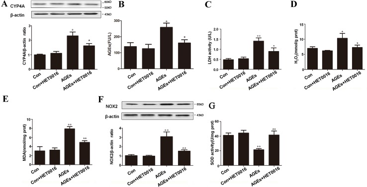
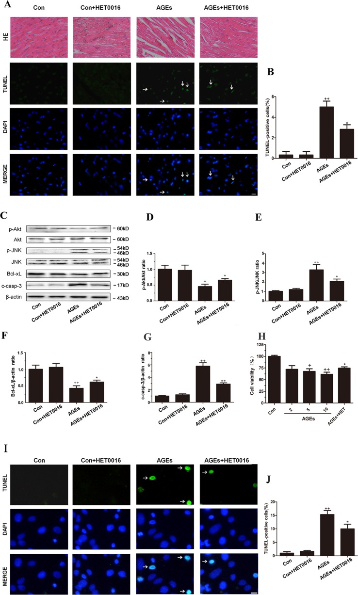
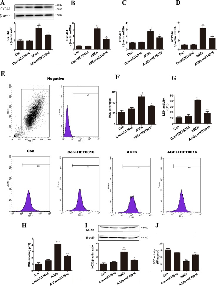
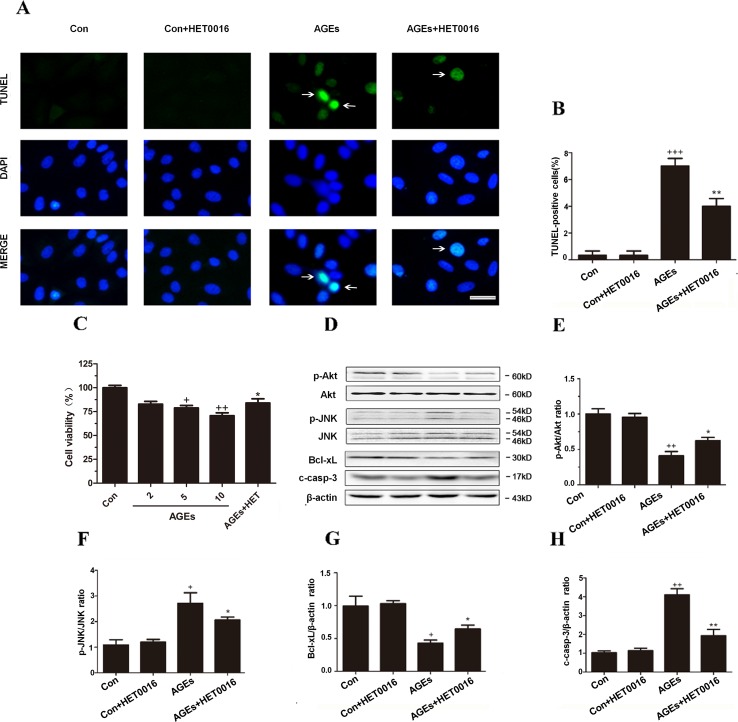
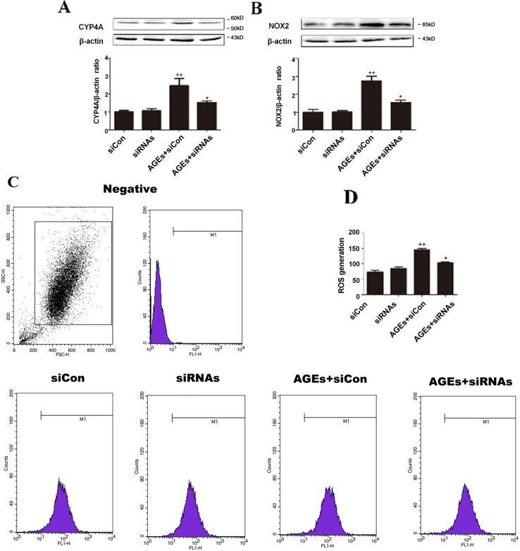
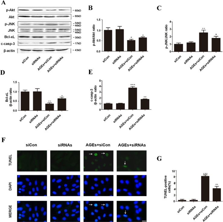
High exposure to advanced glycation end-products (AGEs) may induce cardiotoxicity. However, the effects and mechanisms remain to be further clarified. CYP4A plays an important role in the pathophysiological process of myocardial abnormalities by modulating oxidative stress and apoptosis (OS/Apop) signaling pathway. The present work aimed to investigate whether CYP4A mediates AGEs-induced myocardial injury. AGEs solution was administered intragastrically to C57BL/6 mice for 60 days, while the specific inhibitor of CYP4A, HET0016, was given from the 47th day via intraperitoneal injection for 2 weeks. Levels of OS/Apop in heart tissue were measured. The effects on the cell viability and apoptosis were detected in primary rat cardiomyocytes. To further investigate the mechanism, H9c2 cells were treated with HET0016 or small interfering RNAs (siRNAs) against CYP4a mRNA before incubation with AGEs. Exposure to AGEs led to significantly increased expression of CYP4A and levels of OS/Apop in heart and H9c2 cells both in vivo and in vitro. The OS/Apop pathway was activated with increased expression of NOX2, p-JNK, and cleaved caspase-3 (c-caspase-3) and decreased expression of p-Akt and Bcl-xL both in vivo and in vitro. Specific CYP4A suppression by HET0016 or siRNA exerted significant protective effects by attenuating AGEs-induced OS/Apop pathways in vitro. Our results demonstrate that specific inhibition of CYP4A might be a potential therapeutic option for myocardial injury induced by AGEs.

Keywords: CYP4A; advanced glycation end-products; apoptosis; myocardium; oxidative stress.


Figures

Figure 1
Figure 1
Inhibition of CYP4A with HET0016 reduces myocardial oxidative stress induced by advanced glycation end-products (AGEs) in mice. (A) Protein expression of CYP4A, n = 3. (B) Serum AGEs levels, n = 9. (C) Serum lactate dehydrogenase (LDH) levels, n = 9. (D) H2O2 content in the myocardium, n = 9. (E) Myocardial malondialdehyde (MDA) content, n = 9. (F) Protein expression of NOX2, n = 3. (G) Activity of SOD in the myocardium, n = 9. AGEs solution was administered intragastrically to C57BL/6 mice for 60 days, while the specific inhibitor of CYP4A, HET0016, was given from the 47th day via intraperitoneal injection. Con, control. Compared with the Con group, +p < 0.05, ++p < 0.01; compared with the AGEs group, *p < 0.05, **p < 0.01 (one-way ANOVA with Tukey post hoc).
Figure 2
Figure 2
Inhibition of CYP4A with HET0016 reduces myocardial apoptosis induced by AGEs in mice and cardiomyocytes. (A) Hematoxylin and eosin (HE) staining and TUNEL staining of mouse heart tissues. Scale bar = 50 μm. (B) Relative apoptosis rates are represented as TUNEL-positive cells/DAPI-positive cells, n = 6. (C–G) Protein expression of p-Akt, p-JNK, Bcl-xL, and c-caspase-3. n = 3. (H) Cell viability of rat cardiomyocytes detected with cell counting kit 8. n = 6. (I) Apoptosis of rat cardiomyocytes via TUNEL staining. (J) Relative apoptosis rates are represented as TUNEL-positive cells/DAPI-positive cells, n = 3. Cardiomyocytes were isolated and cultured from ventricles of neonatal rats. Scale bar = 10 μm. AGEs solution was administered intragastrically to C57BL/6 mice for 60 days, while the specific inhibitor of CYP4A, HET0016, was given from the 47th day via intraperitoneal injection. Con, control. Compared with the Con group, +p < 0.05, ++p < 0.01, +++p < 0.001; compared with the AGEs group, *p < 0.05, **p < 0.01 (one-way ANOVA with Tukey post hoc).
Figure 3
Figure 3
Inhibition of CYP4A with HET0016 protects H9c2 cells from AGEs-induced oxidative stress. (A) Protein expression of CYP4A, n = 3. (B–D) mRNA expressions of CYP4a1, CYP4a2, CYP4a3, n = 3. (E) Level of intracellular ROS generation. (F) A summary of flow cytometry analyses of cells stained with 2,7-dichlorodihydrofluoresceindiacetate (DCFH-DA), n = 3. (G) LDH activity in the medium, n = 6. (H) Content of MDA, n = 6. (I) Protein expression of NOX2, n = 3. (J) Activity of SOD, n = 6. H9c2 cells at a density of 1 × 10⁵ cells per well were seeded on six-well plates and treated at the time of 90% confluence. HET0016 or vehicle in fetal bovine serum (PBS)-free media (2 μM) was added 1 h before exposure to 10 μM AGEs or bovine serum albumin (BSA) for 24 h. Con, control. Compared with the Con group, ++p < 0.01, +++p < 0.001; compared with the AGEs group, *p < 0.05, **p < 0.01 (one-way ANOVA with Tukey post hoc).
Figure 4
Figure 4
Inhibition of CYP4A with HET0016 protects H9c2 cells from AGEs-induced apoptosis. (A) Apoptosis via TUNEL staining. Scale bar = 10 μm. (B) Relative apoptosis rates are represented as TUNEL-positive cells/DAPI-positive cells, n = 3. (C) Cell viability detected with cell counting kit 8. n = 10. (DH) Protein expression of p-Akt, p-JNK, Bcl-xL, and c-caspase-3, n = 3. H9c2 cells at a density of 1 × 10⁵ cells per well were seeded on six-well plates and treated at the time of 90% confluence. HET0016 or vehicle in FBS-free media (2 μM) was added 1 h before exposure to 10 μM AGEs or BSA for 24 h. Con, control. Compared with the Con group, +p < 0.05, ++p < 0.01, +++p < 0.001; compared with the AGEs group, *p < 0.05, **p < 0.01 (one-way ANOVA with Tukey post hoc).
Figure 5
Figure 5
Inhibition of CYP4A by siRNAs protects H9c2 cells from AGEs-induced oxidative stress. (A) Protein expression of CYP4A. (B) Protein expression of NOX2. (C) Level of ROS generation. (D) A summary of flow cytometry analyses of cells stained with DCFH-DA. n = 3. siCon: siControl. siRNAs: siRNAs against CYP4a1, CYP4a2, and CYP4a3. H9c2 cells at a density of 1 × 10⁵ cells per well were seeded on six-well plates and transfected at 70–80% confluence with siRNAs against rat CYP4a1, CYP4a2, CYP4a3, or nonbinding control siRNA. Transfection was performed with Lipofectamine 2000. After 24 h of Cyp4a siRNAs transfection, cells were treated with 10 μM AGEs or BSA for 24 h. Compared with the siCon group, ++p < 0.01; compared with the AGEs+siCon group, *p < 0.05, **p < 0.01 (one-way ANOVA with Tukey post hoc).
Figure 6
Figure 6
Inhibition of CYP4A by siRNAs protects H9c2 cells from AGEs-induced apoptosis. (AE) Protein expression of p-Akt, p-JNK, Bcl-xL, and c-caspase-3, n = 3. (F) Relative apoptosis rates are represented as TUNEL-positive cells/DAPI-positive cells, n = 6. (G) Apoptosis via TUNEL staining. H9c2 cells at a density of 1 × 10⁵ cells per well were seeded on six-well plates and transfected at 70–80% confluence with siRNAs against rat CYP4a1, CYP4a2, CYP4a3, or nonbinding control siRNA. Transfection was performed with Lipofectamine 2000. After 24 h of CYP4a siRNAs transfection, cells were treated with 10 μM AGEs or BSA for 24 h. Scale bar = 10 μm. siCon: siControl. siRNAs: siRNAs against CYP4a1, CYP4a2, and CYP4a3. Compared with the siCon group, + p < 0.05, ++ p < 0.01, +++ p < 0.001; compared with the AGEs+siCon group, *p < 0.05, **p < 0.01 (one-way ANOVA with Tukey post hoc).

References

    1. Adamopoulos C., Piperi C., Gargalionis A. N., Dalagiorgou G., Spilioti E., Korkolopoulou P., et al. (2016). Advanced glycation end products upregulate lysyl oxidase and endothelin-1 in human aortic endothelial cells via parallel activation of ERK1/2-NF-kappaB and JNK-AP-1 signaling pathways. Cell. Mol. Life Sci. 73 (8), 1685–1698. 10.1007/s00018-015-2091-z - DOI - PMC - PubMed
    1. Alsaad A. M., Zordoky B. N., Tse M. M., El-Kadi A. O. (2013). Role of cytochrome P450-mediated arachidonic acid metabolites in the pathogenesis of cardiac hypertrophy. Drug Metab. Rev. 45 (2), 173–195. 10.3109/03602532.2012.754460 - DOI - PubMed
    1. Bodiga V. L., Eda S. R., Bodiga S. (2014). Advanced glycation end products: role in pathology of diabetic cardiomyopathy. Heart Fail. Rev. 19 (1), 49–63. 10.1007/s10741-013-9374-y - DOI - PubMed
    1. Cai W., Uribarri J., Zhu L., Chen X., Swamy S., Zhao Z., et al. (2014). Oral glycotoxins are a modifiable cause of dementia and the metabolic syndrome in mice and humans. Proc. Natl. Acad. Sci. U.S.A. 111 (13), 4940–4945. 10.1073/pnas.1316013111 - DOI - PMC - PubMed
    1. Chen M., Li H., Wang G., Shen X., Zhao S., Su W. (2016). Atorvastatin prevents advanced glycation end products (AGEs)-induced cardiac fibrosis via activating peroxisome proliferator-activated receptor gamma (PPAR-gamma). Metabolism 65 (4), 441–453. 10.1016/j.metabol.2015.11.007 - DOI - PubMed