. 2019 Aug 6:10:1800.
doi: 10.3389/fmicb.2019.01800. eCollection 2019.

Dependence on Dectin-1 Varies With Multiple Candida Species

Affiliations

Dependence on Dectin-1 Varies With Multiple Candida Species

Aiysha Thompson et al. Front Microbiol. .

Abstract

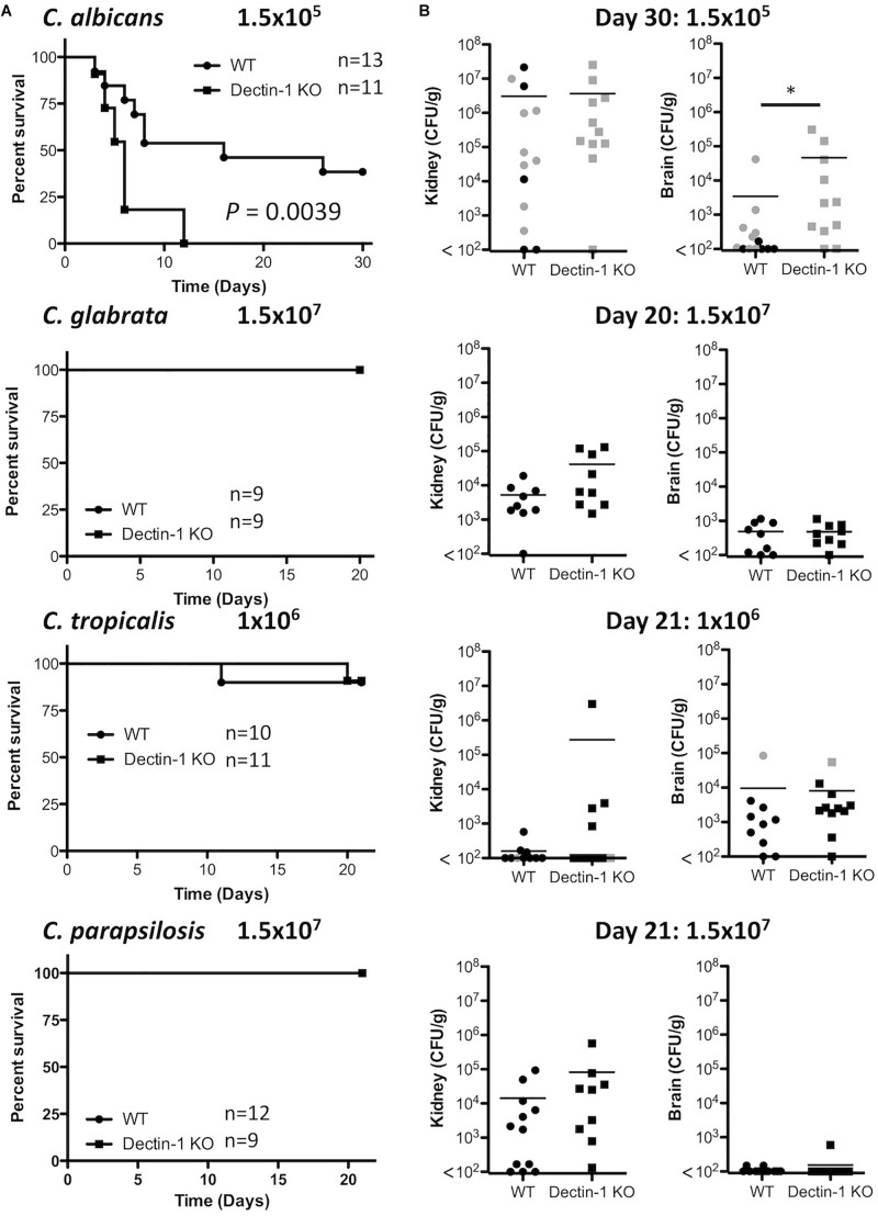
Four Candida spp. (albicans, glabrata, tropicalis, parapsilosis) cause >95% of invasive Candida infections. C. albicans elicits immune responses via pathogen recognition receptors including C-type lectin-like receptors (CLRs). The CLR, Dectin-1 is important for host immunity to C. albicans and C. glabrata, however, whether Dectin-1 is important for host defense against C. tropicalis or C. parapsilosis is unknown. Therefore, we compared the involvement of Dectin-1 in response to these four diverse Candida spp. We found that Dectin-1 mediates innate cytokine responses to these Candida spp. in a species- and cell-dependent manner. Dectin-1 KO mice succumbed to infection with highly virulent C. albicans while they mostly survived infection with less virulent Candida spp. However, Dectin-1 KO mice displayed increased fungal burden following infection with each Candida spp. Additionally, T cells from Dectin-1 KO mice displayed enhanced effector functions likely due to the inability of Dectin-1 KO mice to clear the infections. Together, these data indicate that Dectin-1 is important for host defense to multiple Candida spp., although the specific roles for Dectin-1 varies with different Candida spp.

Keywords: Candida spp.; Dectin-1; T cells; dendritic cells; macrophages.


Figures

FIGURE 1
FIGURE 1
Candida spp. display differential β1,3-glucan exposure. (A,B) C. albicans SC5314, C. glabrata SCS74761, C. tropicalis SCS74663 and C. parapsilosis SCSB5882 were incubated with Dectin-1 Fc, Micl Fc or PBS followed by incubation with Pe-conjugated Donkey anti-human IgG. β1,3-glucan exposure on the Candida cell surface was analyzed by flow cytometry. (A) Representative flow plots display β1,3-glucan exposure from 3 independent experiments. (B) Graphs display MFI of control Micl Fc or Dectin-1 Fc minus MFI of Pe-conjugated Donkey anti-human IgG. Graphs display cumulative data from 3 independent experiments. 2-way ANOVA with Bonferroni’s post-test I = Interaction between Candida spp. and CLR Fc binding.
FIGURE 2
FIGURE 2
Dectin-1 mediates cytokine responses to multiple Candida spp. BMDCs (A,E) and BMDMs (B,F) from WT and Dectin-1 KO mice were stimulated with C. albicans SC5314 (Ca), C. glabrata SCS74761 (Cg), C. tropicalis SCS74663 (Ct) and C. parapsilosis SCSB5882 (Cp) at a ratio of 1:1 Candida:Cells. (C) BMDMs from WT and Dectin-1 KO mice were stimulated with C. albicans AM2005/0463, C. glabrata SCSB5311, C. tropicalis AM2007/0112 and C. parapsilosis AM2005/0207 at a ratio of 1:1 Candida:Cells. (D) BMDMs from WT and Dectin-1 KO mice were stimulated with thimerosal-killed C. albicans SC5314 (TK Ca), C. glabrata SCS74761 (TK Cg), C. tropicalis SCS74663 (TK Ct) and C. parapsilosis SCSB5882 (TK Cp) at a ratio of 1:1 Candida:Cells. Cytokine levels (A–D) and nitric oxide levels (E,F) in the supernatants were measured after 24h incubation. Results are presented as means ± s.e.m. of 5–6 (A), 4 (B), 3 (C), 2–4 (D), 3 (E) or 4 (F) independent experiments. (2-way ANOVA, Bonferroni’s post-test) I = Interaction, G = Genotype, C = Candida.
FIGURE 3
FIGURE 3
Dectin-1 KO mice are susceptible to infection with highly virulent C. albicans. (A) WT and Dectin-1 KO mice were infected intravenously with the indicated doses of C. albicans for 30 days, C. glabrata for 20 days, C. tropicalis for 21 days or C. parapsilosis for 21 days. Survival curves based on humane end-point of infected WT (filled circles) and Dectin-1 KO mice (filled squares). Graphs are the cumulative result of 2 independent experiments. Log-rank test. (B) CFU in the kidneys at 20–30 days after infection (black symbols) or at time of death by humane end point (gray symbols). Graphs are the cumulative result of 2 independent experiments. Each symbol represents an individual mouse. Student’s t test on transformed data.
FIGURE 4
FIGURE 4
Dectin-1 mediates Candida clearance. (A,B) WT and Dectin-1 KO mice were infected intravenously with the indicated doses of C. albicans, C. glabrata, C. tropicalis or C. parapsilosis for 7 days. (A) CFU in the kidneys and brains of mice at 7 days post-infection were determined. Graphs are the cumulative result of 2–4 independent experiments. Each symbol represents an individual mouse. Student’s t test (C. albicans: transformed data) (B) Cytokine levels in the serum of mice at 7 days post-infection. Graphs are the cumulative result of 2–4 independent experiments. Each symbol represents an individual mouse. Student’s t test/ Mann-Whitney test (IL-6, TNF, IL-1β, IL-10: transformed data).
FIGURE 5
FIGURE 5
Dectin-1 regulates Candida-associated T cell responses. (A–C) WT and Dectin-1 KO mice were intravenously infected with 1.5 × 10⁴ CFU C. albicans, 1.5 × 10⁷ CFU C. glabrata, 1 × 10⁶ CFU C. tropicalis or 1.5 × 10⁷ CFU C. parapsilosis for 7 days. (A) RNA was isolated from the spleens of infected mice, cDNA was prepared and mRNA transcripts were detected by real-time qPCR. mRNA levels were normalized to Hprt1. Graphs display the cumulative result of 2 independent experiments. Each symbol represents one mouse. (Student’s t test) (B) Splenocytes from infected mice were restimulated with PMA/Ionomycin for 4 h and IFN-γ and IL-17 levels were measured by FACS. Graphs display% T cells producing IFN-γ - Isotype or MFI of IL-17 production by T cells. IL-17 MFI by cells from one WT mouse stimulated with PMA/Ionomycin was set at 100% and all other IL-17 MFI in that experiment are presented as a% of this. MFI for the Isotype was then subtracted. (C) Splenocytes from infected mice were restimulated with live Candida spp. for 48 h and IFN-γ and IL-17 levels were measured by ELISA. (A–C) Graphs are the cumulative result of 2–4 independent experiments. Each symbol represents an individual mouse. Student’s t test/Mann-Whitney test.

References

    1. Alangaden G. J. (2011). Nosocomial fungal infections: epidemiology, infection control, and prevention. Infect. Dis. Clin. North Am. 25 201–225. 10.1016/j.idc.2010.11.003 - DOI - PubMed
    1. Anaissie E., Hachem R., K-Tin-U C., Stephens L. C., Bodey G. P. (1993). Experimental hematogenous candidiasis caused by Candida krusei and Candida albicans: species differences in pathogenicity. Infect. Immun. 61 1268–1271. - PMC - PubMed
    1. Arendrup M., Horn T., Frimodt-Moller N. (2002). In vivo pathogenicity of eight medically relevant Candida species in an animal model. Infection 30 286–291. 10.1007/s15010-002-2131-0 - DOI - PubMed
    1. Ballou E. R., Avelar G. M., Childers D. S., Mackie J., Bain J. M., Wagener J., et al. (2016). Lactate signalling regulates fungal beta-glucan masking and immune evasion. Nat. Microbiol. 2:16238. 10.1038/nmicrobiol.2016.238 - DOI - PMC - PubMed
    1. Branzk N., Lubojemska A., Hardison S. E., Wang Q., Gutierrez M. G., Brown G. D., et al. (2014). Neutrophils sense microbe size and selectively release neutrophil extracellular traps in response to large pathogens. Nat. Immunol. 15 1017–1025. 10.1038/ni.2987 - DOI - PMC - PubMed