. 2019 Aug 7:10:704.
doi: 10.3389/fgene.2019.00704. eCollection 2019.

Copy Number Variation of Human Satellite III (1q12) With Aging

Affiliations

Copy Number Variation of Human Satellite III (1q12) With Aging

Elizaveta S Ershova et al. Front Genet. .

Abstract

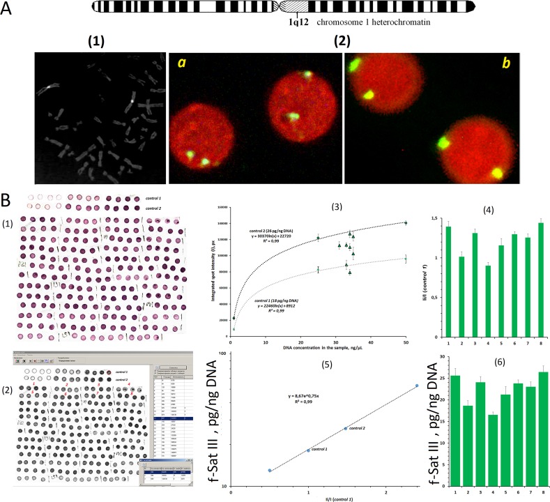
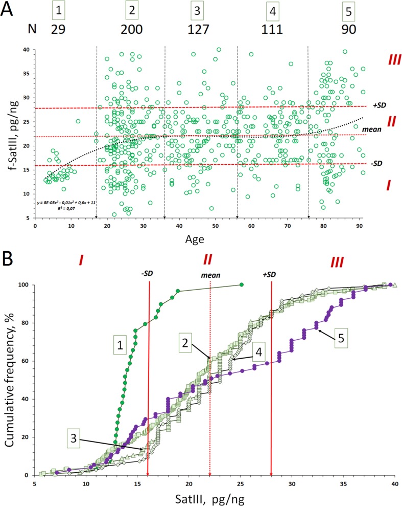
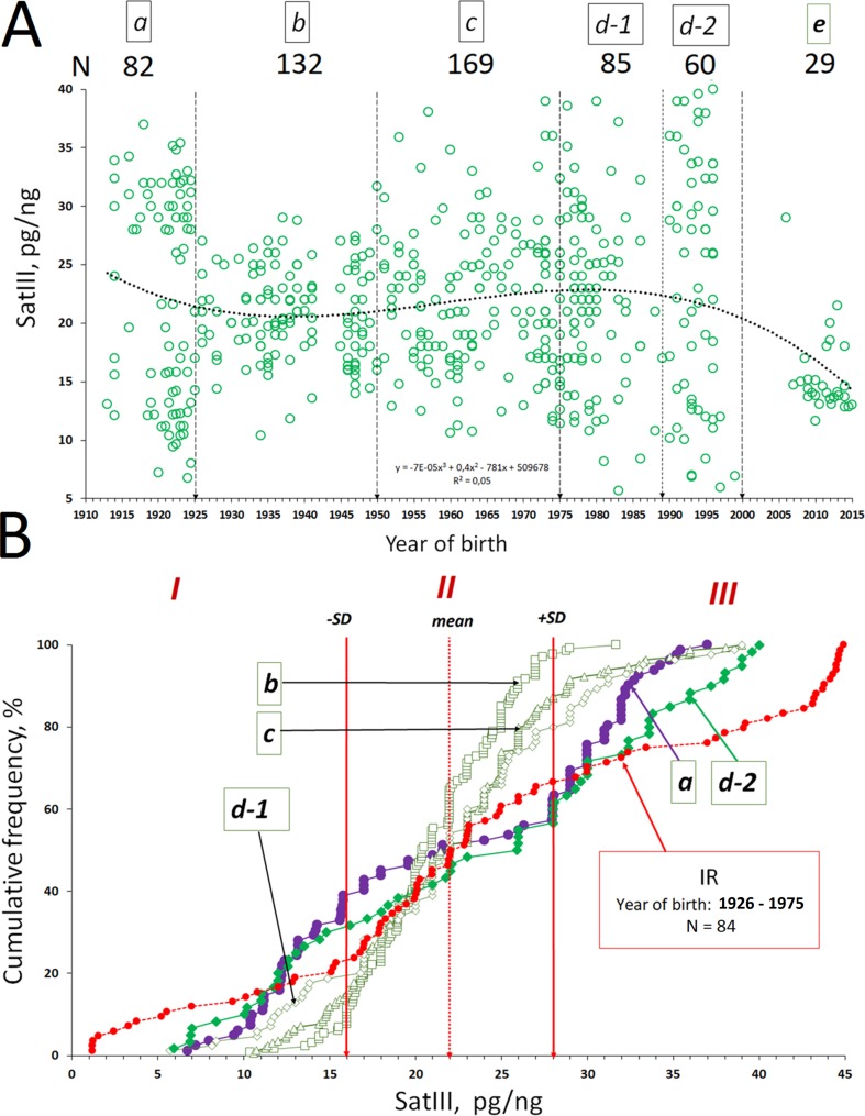
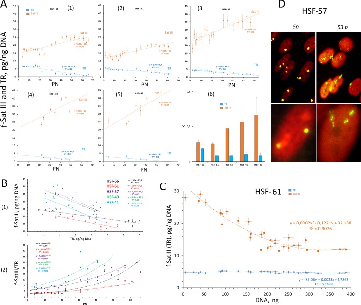
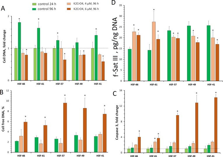
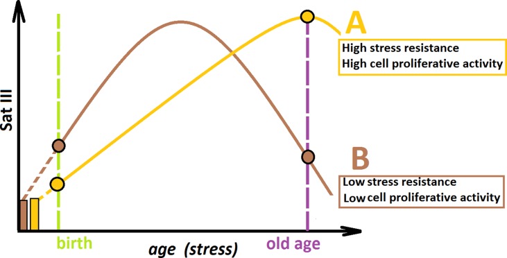
Introduction: Human satellite DNA is organized in long arrays in peri/centromeric heterochromatin. There is little information about satellite copy number variants (CNVs) in aging and replicative cell senescence (RS). Materials and Methods: Biotinylated pUC1.77 probe was used for the satellite III (f-SatIII) quantitation in leukocyte DNA by the non-radioactive quantitative hybridization for 557 subjects between 2 and 91 years old. The effect of RS and genotoxic stress (GS, 4 or 6 µM of K2CrO4) on the f-SatIII CNV was studied on the cultured human skin fibroblast (HSF) lines of five subjects. Results: f-SatIII in leukocyte and HSFs varies between 5.7 and 40 pg/ng of DNA. During RS, the f-SatIII content in HSFs increased. During GS, HSFs may increase or decrease f-SatIII content. Cells with low f-SatIII content have the greatest proliferative potential. F-SatIII CNVs in different individuals belonging to the different generations depend on year of their birth. Children (born in 2005-2015 years) differed significantly from the other age groups by low content and low coefficient of variation of f-SatIII. In the individuals born in 1912-1925 and living in unfavorable social conditions (FWW, the Revolution and the Russian Civil War, SWW), there is a significant disproportion in the content of f-SatIII. The coefficient of variation reaches the maximum values than in individuals born in the period from 1926 to 1975. In the group of people born in 1990-2000 (Chernobyl disaster, the collapse of the Soviet Union, and a sharp decline in the population living standard), again, there is a significant disproportion of individuals in the content of f-SatIII. A similar disproportion was observed in the analysis of a group of individuals born in 1926-1975 who in their youth worked for a long time in high-radioactive environment. Conclusion: In generations that were born and who lived in childhood in a period of severe social perturbations or in conditions of environmental pollution, we found a significant increase in leukocyte DNA f-SatIII variability. It is hypothesized that the change of the f-SatIII content in the blood cells reflects the body response to stress of different nature and intensity.

Keywords: aging; copy number variance; genotoxic stress response; replicative senescence; satellite DNA.


Figures

Figure 1
Figure 1
Determination of f-SatIII in human leukocyte DNA using non-radioactive quantitative hybridization (NQH). (A) (1) The hybridization of bio-pUC1.77 probe specifically to the lql2 region of the human chromosome 1 (f-SatIII). (2) The hybridization of the bio-pUC1.77 with the nuclei of the human lymphocytes: (a) a 17-year-old donor (6.7 pg of f-SatIII/ng DNA, detected with NQH); (b) a 77-year-old donor (31 pg of f-SatIII/ng DNA). The figures (1 and 2) show the FISH data obtained earlier in our work (Ermakov et al., 2009). (B) (1) Photo of the membrane fragments with visualized f-SatIII. Four spots are applied for each DNA sample. Besides, standard genomic DNA samples (control 1 and control 2) were applied onto the same filter in order to define the calibration dependence of the signal on the number of the repeats in the sample. The DNA concentration in the standard calibration sample ranged within 5–50 ng/µl. (2) The filter was scanned, and the average integral intensity I of the spots was determined. (3) Dependence of I on DNA concentration in the sample plotted for the standard samples (dashed line) and for tested DNA samples 1–8. (4) The ratio I i/I control1 was calculated. (5) Calibration dependence of f-SatIII on I i/I control1 ratio plotted for four DNA samples with known f-SatIII content. (6) The f-SatIII content in the genome was calculated. This experiment is detailed in the Supplement .
Figure 2
Figure 2
Copy number variants (CNVs) of f-SatIII in the human blood leukocyte DNA samples with age. (A) Association of the f-SatIII content with the age for the 557 subjects aged 2 to 91 years. The graph shows the area with low (I), moderate (II), and high (III) f-SatIII contents. All the individuals were divided into five age groups ( Table 1A ). (B) Cumulative distribution of the f-SatIII content in groups 1–5 of individuals of different age. Distributions for children (group 1) and for the elderly (group 5) are significantly different from those of the other groups ( Table 1B ).
Figure 3
Figure 3
The dependence of the f-SatIII CNV in human leukocytes from a person’s year of birth. (A) Association of the f-SatIII content with a person’s year of birth for the 557 subjects. The total group of the adults who were born in the 20th century was divided into five groups (a–d, Table 2A ). (B) Cumulative distribution of the f-SatIII content in groups a–d. Red line (group IR) shows f-SatIII content in the genomes of the adults who were born in 1926–1975. These individuals have worked for a long time with ionizing radiation sources. We published these data earlier (Korzeneva et al., 2016). Distributions for group a (1912–1925 year of birth), group d-2 (1990–2000), and IR group (1926–1975) are significantly different from those of the other groups ( Table 2B ).
Figure 4
Figure 4
Changes in the f-SatIII content during the replicative senescence of the cultured skin fibroblasts (HSFs). (A) (1–5) The dependence of the f-SatIII and telomere repeat (TR) content in the genomes of HSFs on the number of passages (PN). The linear regression data are shown in the graphs. (6) The rate of change in the content of f-SatIII and TR in the genome of the HSFs during the replicative senescence. (B) (1) The dependence of the f-SatIII content in the genomes of HSFs on the TR content. (2) The dependence of the f-SatIII/TR ratio in the genomes of HSFs on the number of passages. (C) The dependence of the f-SatIII and TR content in the genomes of HSF-61 on the total cell DNA content. The suspension of the cells (fifth passage) was seeded into the wells in terms of not more than 10–20 cells per well. Cells were cultured for 3 weeks. DNA was isolated from these wells, and its amount was measured. (D) The hybridization of pUC1.77 probe (f-SatIII) labeled with biotin to the lql2 region of the human chromosome 1 in HSF-57 (early and late passages). The data were received and kindly provided by M.S. Konkova.
Figure 5
Figure 5
HSF response to genotoxic effect of K2CrO4. Cells were seeded onto a 24-well plate and cultured for 48 h. Then 4 or 6 µM of K2CrO4 was added, cells were incubated for 24 h (point 24 h), the medium was replaced, and incubation was continued for up to 72 h (point 96 h). Cells of three to six wells were examined for each K2CrO4 concentration and each control variant. (A) Change for the cell DNA on the plate. (B) Change for the cell-free DNA in the medium. (C) Change in caspase 3 activity in the protein lysate of the cells. (D) Change in the f-SatIII content in the DNA. *p < 0.05.
Figure 6
Figure 6
A hypothetical scheme illustrating the changes in the f-SatIII content during human aging. Individual (A) and individual (B) have the same f-SatIII repeat copy number at the time of conception. (A) is more resistant to genotoxic stress than (B). During an aging under identical living conditions, the content of f-SatIII increases faster in (B). At the young age, the f-SatIII content in B will be higher. Reaching its maximum, the f-SatIII content of (B) will start to decline, as aging is associated with increased stress and the cells with high f-SatIII content die. If (B) would live to an old age, the f-SatIII content analysis will reveal high values for (A) and low values for (B). The proportion of the individual with high f-SatIII content in the elderly group is significantly increased. Therefore, it seems that the chances for a longer life are greater for (A).

References

    1. Asatiani N., Kartvelishvili T., Abuladze M., Asanishvili L., Sapojnikova N. (2011). Chromium (VI) can activate and impair antioxidant defense system. Biol. Trace Elem. Res. 142 (3), 388–397. 10.1007/s12011-010-8806-y - DOI - PubMed
    1. Bersani F., Lee E., Kharchenko P. V., Xu A. W., Liu M., Xega K., et al. (2015). Pericentromeric satellite repeat expansions through RNA-derived DNA intermediates in cancer. Proc. Natl. Acad. Sci. U. S. A. 112 (49), 15148–15153. 10.1073/pnas.1518008112 - DOI - PMC - PubMed
    1. Bierhoff H., Postepska-Igielska A., Grummt I. (2014). Noisy silence: non-coding RNA and heterochromatin formation at repetitive elements. Epigenetics 9 (1), 53–61. 10.4161/epi.26485 - DOI - PMC - PubMed
    1. Black E. M., Giunta S. (2018). Repetitive fragile sites: centromere satellite DNA as a source of genome instability in human diseases. Genes (Basel) 9 (12), E615. 10.3390/genes9120615 - DOI - PMC - PubMed
    1. Brahmachary M., Guilmatre A., Quilez J., Hasson D., Borel C., Warburton P., et al. (2014). Digital genotyping of macrosatellites and multicopy genes reveals novel biological functions associated with copy number variation of large tandem repeats. PLoS Genet. 10 (6), e1004418. 10.1371/journal.pgen.1004418 - DOI - PMC - PubMed

LinkOut - more resources