. 2018 Nov 28;6(44):7185-7196.
doi: 10.1039/C8TB01116H. Epub 2018 Jun 18.

Conductive Silk-Polypyrrole Composite Scaffolds with Bioinspired Nanotopographic Cues for Cardiac Tissue Engineering

Affiliations

Conductive Silk-Polypyrrole Composite Scaffolds with Bioinspired Nanotopographic Cues for Cardiac Tissue Engineering

Jonathan H Tsui et al. J Mater Chem B. .

Abstract

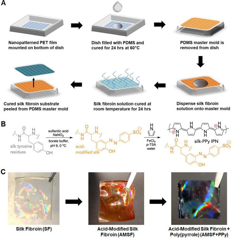
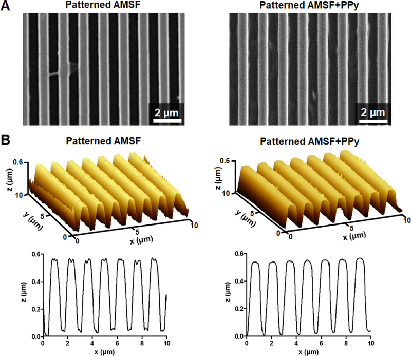
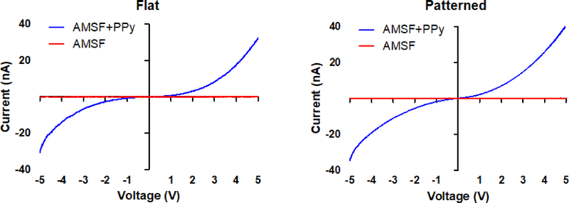
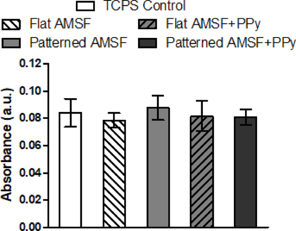
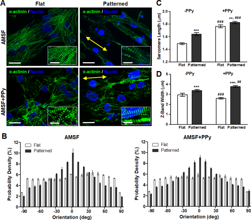
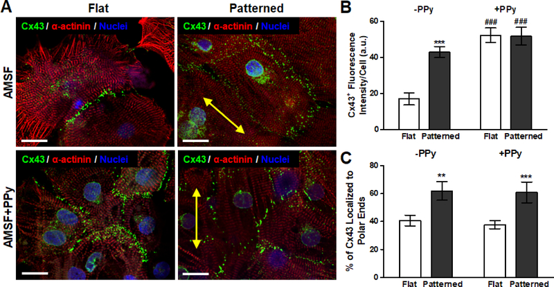
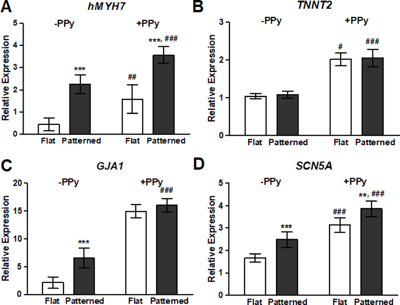
We report on the development of bioinspired cardiac scaffolds made from electroconductive acid-modified silk fibroin-poly(pyrrole) (AMSF+PPy) substrates patterned with nanoscale ridges and grooves reminiscent of native myocardial extracellular matrix (ECM) topography to enhance the structural and functional properties of cultured human pluripotent stem cells (hPSC)-derived cardiomyocytes. Nanopattern fidelity was maintained throughout the fabrication and functionalization processes, and no loss in conductive behavior occurred due to the presence of the nanotopographical features. AMSF+PPy substrates were biocompatible and stable, maintaining high cell viability over a 21-day culture period while displaying no signs of PPy delamination. The presence of anisotropic topographical cues led to increased cellular organization and sarcomere development, and electroconductive cues promoted a significant improvement in the expression and polarization of connexin 43 (Cx43), a critical regulator of cell-cell electrical coupling. The combination of biomimetic topography and electroconductivity also increased the expression of genes that encode key proteins involved in regulating the contractile and electrophysiological function of mature human cardiac tissue.

Keywords: cardiac tissue engineering; electroconductivity; nanotopography; polypyrrole; silk fibroin; stem cell-derived cardiomyocyte.


Conflict of interest statement

Conflicts of Interest Deok-Ho Kim is a co-founder and scientific board member at NanoSurface Biomedical Inc.

Figures

Figure 1.
Figure 1.. Fabrication of nanopatterned acid-modified silk fibroin and polypyrrole deposition.
(A) Scheme of the process utilized to generate the master mold and subsequent casting of silk fibroin solution to generate nanopatterned substrates. (B) Description of chemical moieties and reactions that occur during the acid modification and PPy deposition stages. (C) Representative photographs of the nanopatterned silk substrates at each stage. The substrates take on an orange hue after acid modification and turn black after PPy deposition. An iridescent sheen is present throughout. Dimensions of the pictured films are 1 cm × 1 cm.
Figure 2.
Figure 2.. Characterization of substrate topography.
(A) Representative SEM images of nanopatterned substrates with and without PPy. (B) 3D and cross-section AFM profiles illustrate that the fidelity of the nanotopography is well-maintained even after PPy deposition.
Figure 3.
Figure 3.. AMSF+PPy substrates are electrically responsive.
I-V curves of flat and patterned substrates indicate that the electroconductive property of the substrates is unaffected by topographical changes.
Figure 4.
Figure 4.. AMSF and AMSF+PPy substrates are biocompatible.
Colorimetric MTS assays of cardiomyocytes cultured for 21 days show that acid-modification and the presence of PPy have no negative impact on cell viability. Viability is presented as normalized absorbance values.
Figure 5.
Figure 5.. Nanotopographical and electroconductive cues enhance structural organization and contractile apparatus development.
(A) Representative images of cardiomyocytes fluorescently stained for α-actinin (green) and nuclei (blue). Cells on nanopatterned substrates exhibit elongated and aligned morphologies. Yellow arrows indicate the direction of the nanopattern. Scale bars: 25 μm; inset scale bars: 10 μm. (B) Anisotropic nanopatterns induce cell orientation along the axis of the pattern, indicated here by 0°, whereas cells are randomly oriented on flat substrates. (C) Sarcomere length is significantly increased due to nanopatterning and PPy. (D) Z-band width is greatly influenced by topography but is only slightly increased on nanopatterned substrates due to PPy. All quantitative data are presented as means ± SEM, n≥10 different cultures. **p < 0.01, ***p < 0.001 (flat vs. patterned; Student’s t-test); ##p < 0.01, ###p < 0.001 (−PPy vs. +PPy; Student’s t-test).
Figure 6.
Figure 6.. Modulation of gap junction protein expression and localization due to electroconductivity and anisotropic nanotopography.
(A) Representative images of cardiomyocytes fluorescently stained for α-actinin (red), Cx43 (green), and nuclei (blue). Yellow arrows indicate the direction of the nanopattern. Scale bars: 25 μm. (B) While bioinspired nanotopography significantly increased Cx43 expression, the advantage of topographical cues is nearly nullified in the presence of electroconductive cues as PPy elicited a similar degree of Cx43 expression on both flat and patterned substrates. (C) Cx43 polarization is not significantly affected by electroconductive cues as topography dictated protein localization patterns. All quantitative data are presented as means ± SEM, n≥10 different cultures. **p < 0.01, ***p < 0.001 (flat vs. patterned; Student’s t-test); ###p < 0.001 (-PPy vs. +PPy; Student’s t-test).
Figure 7.
Figure 7.. Nanotopography and electroconductivity synergistically induced the largest increases in genetic markers for cardiac maturation.
qRT-PCR analysis of the relative expression levels of: (A) β-myosin heavy chain (hMYH7), (B) cardiac troponin T2 (TNNT2), (C) connexin 43 (GJA1), and (D) NaV1.5 (SCN5A). Presence of biomimetic nanotopography significantly increased gene expression, with the exception of TNNT2. Cardiomyocytes cultured on electroconductive silk substrates exhibited an increased expression of all assayed genes when compared to cells on non-conductive substrates. All data are presented as means ± SEM, n≥6 different cultures, **p < 0.01, ***p < 0.001 (flat vs. patterned; Student’s t-test); #p < 0.05, ##p < 0.01, ###p < 0.001 (-PPy vs. +PPy; Student’s t-test).

References

    1. Guo BL, Glavas L and Albertsson AC, Prog Polym Sci, 2013, 38, 1263–1286.
    1. Lee TJ, Park S, Bhang SH, Yoon JK, Jo I, Jeong GJ, Hong BH and Kim BS, Biochem. Biophys. Res. Commun, 2014, 452, 174–180. - PubMed
    1. Shin SR, Zihlmann C, Akbari M, Assawes P, Cheung L, Zhang KZ, Manoharan V, Zhang YS, Yuksekkaya M, Wan KT, Nikkhah M, Dokmeci MR, Tang XW and Khademhosseini A, Small, 2016, 12, 3677–3689. - PMC - PubMed
    1. Martinelli V, Cellot G, Toma FM, Long CS, Caldwell JH, Zentilin L, Giacca M, Turco A, Prato M, Ballerini L and Mestroni L, ACS Nano, 2013, 7, 5746–5756. - PubMed
    1. Shin SR, Jung SM, Zalabany M, Kim K, Zorlutuna P, Kim SB, Nikkhah M, Khabiry M, Azize M, Kong J, Wan KT, Palacios T, Dokmeci MR, Bae H, Tang XW and Khademhosseini A, ACS Nano, 2013, 7, 2369–2380. - PMC - PubMed

Publication types

MeSH terms