. 2019 Aug 6:7:190.
doi: 10.3389/fbioe.2019.00190. eCollection 2019.

Laminin-Coated Electrospun Regenerated Silk Fibroin Mats Promote Neural Progenitor Cell Proliferation, Differentiation, and Survival in vitro

Affiliations

Laminin-Coated Electrospun Regenerated Silk Fibroin Mats Promote Neural Progenitor Cell Proliferation, Differentiation, and Survival in vitro

Guangfei Li et al. Front Bioeng Biotechnol. .

Abstract

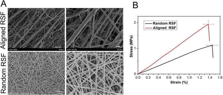
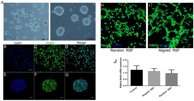
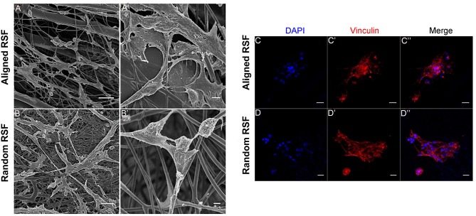
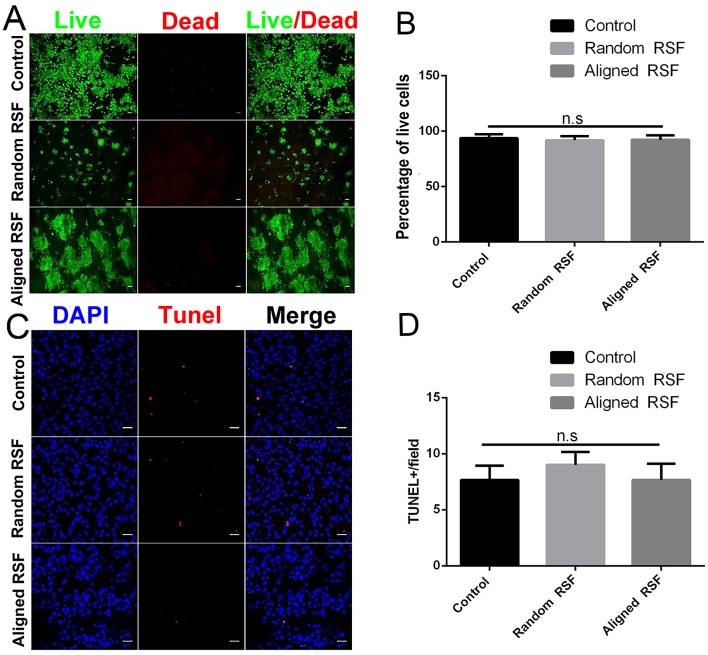
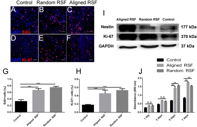
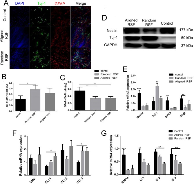
Neural progenitor cell (NPC) transplantation is a promising technique for central nervous system (CNS) reconstruction and regeneration. Biomaterial scaffolds, frameworks, and platforms can support NPC proliferation and differentiation in vitro as well as serve as a temporary extracellular matrix after transplantation. However, further applications of biomaterials require improved biological attributes. Silk fibroin (SF), which is produced by Bombyx mori, is a widely used and studied protein polymer for biomaterial application. Here, we prepared aligned and random eletrospun regenerated SF (RSF) scaffolds, and evaluated their impact on the growth of NPCs. First, we isolated NPCs and then cultured them on either laminin-coated RSF mats or conventional laminin-coated coverslips for cell assays. We found that aligned and random RSF led to increases in NPC proliferation of 143.8 ± 13.3% and 156.3 ± 14.7%, respectively, compared to controls. Next, we investigated neuron differentiation and found that the aligned and the random RSF led to increases in increase in neuron differentiation of about 93.2 ± 6.4%, and 3167.1 ± 4.8%, respectively, compared to controls. Furthermore, we measured the survival of NPCs and found that RSF promoted NPC survival, and found there was no difference among those three groups. Finally, signaling pathways in cells cultured on RSF mats were studied for their contributions in neural cell differentiation. Our results indicate that RSF mats provide a functional microenvironment and represent a useful scaffold for the development of new strategies in neural engineering research.

Keywords: biocompatibility; differentiation; neural progenitor cells; proliferation; regenerated silk fibroin mats.


Figures

Figure 1
Figure 1
SEM images (A) and tensile strength (B) of electrospun RSF mats with aligned fibers and randomly collected fibers. Scale bar: 50 μm.
Figure 2
Figure 2
Characterization of NPCs. (A) Neurosphere formation of NPCs at DIV 5. Immunostaining against nestin (green), an NPC marker, in the individual NPCs (B–D) and neurospheres (E–G). Immunostaining against nestin (green) with NPCs on random and aligned RSF mats (H,I). mRNA expression of Nestin in NPCs grown on aligned and random RSF mats and laminin-coating coverslips for 5 days (J). Nuclei were stained with DAPI (blue). Scale bar: 50 μm.
Figure 3
Figure 3
NPC adhesion on random and aligned RSF mats after 3 days of culture in proliferation medium. Low-magnification (A,B) and high-magnification (A,B) SEM images of NPCs cultured on laminin-coated random and aligned RSF mats in proliferation medium. The high-magnification SEM images illustrate the interaction between the cell filopodia and the RSF mat surface. Immunostaining against vinculin in NPC progeny cultured on the aligned (C–C”) and random RSF mats (D–D”) in proliferation medium after 3 days culture. Scale bar: 50 μm.
Figure 4
Figure 4
(A) Cell viability assay of NPCs on random and aligned RSF mats after 5 days of culture as determined by live/dead assay. Live cells are stained green and dead cells are red. Scale bar: 50 μm. (B) The percentage of live cells on the coverslip and the random and aligned RSF mats. (C) Representative photographs of TUNEL staining (which detects DNA fragmentation as an indicator of apoptosis) in NPCs cultured on coverslips and random and aligned RSF mats. All nuclei were counterstained with DAPI in blue, and all TUNEL-positive cells are red. Scale bar: 20 μm. (D) The average number of TUNEL-positive nuclei per field is shown (n = 20 fields for all three substrates).
Figure 5
Figure 5
NPC proliferation on coverslip controls and random and aligned RSF mats after culturing for 7 days. The expression of EdU on control (A), random RSF mats (B), and aligned RSF mats (C) and the respective expression of Ki-67 (D–F). The percentages of EdU+ cells (G) and Ki-67+ cells (H) in the three groups. (I) Western blot analysis of Ki-67 and nestin expression on coverslip controls and random and aligned RSF mats. (J) The histogram depicts the CCK-8 assay on the coverslip controls and RSF mats after culturing for 1, 3, 5, 7 days in proliferation medium, respectively. The data are presented as mean ± standard error of the mean, **p < 0.01, ***p < 0.001. Scale bar: 50 μm.
Figure 6
Figure 6
The differentiation of NPCs on coverslip controls and random and aligned RSF mats after culturing for 7 days. (A) Representative fluorescence images of differentiated NPCs under differentiation conditions. The cells were immunostained with Tuj-1 for neurons (green), GFAP for astrocytes (red), and DAPI for nuclei (blue). The percentages of Tuj-1+ cells (B) and GFAP+ cells (C) among differentiated NPCs in the three groups. (D) Western blot analysis of nestin and Tuj-1 protein expression of differentiated NPCs on coverslip controls and aligned and random RSF mats. (E) qPCR analysis of nestin, Tuj-1, GFAP, and Olig1 mRNA expression in differentiated NPCs cultured on the three substrates. (F) qPCR analysis of the relative mRNA expression of Smo and Gli1–3, which are involved in the SHH signal pathway, after 7 days of differentiation on the three substrates. (G) qPCR analysis of the relative mRNA expression of BMP4 and Id1–3, which are involved in the BMP4 signaling pathway, after 7 days of differentiation on the three substrates. The data are presented as the mean ± standard error of the mean, *p < 0.05, **p < 0.01, ***p < 0.001. Scale bar: 50 μm.

References

    1. Al-Maawi S., Orlowska A., Sader R., James Kirkpatrick C., Ghanaati S. (2017). In vivo cellular reactions to different biomaterials-Physiological and pathological aspects and their consequences. Semin. Immunol. 29, 49–61. 10.1016/j.smim.2017.06.001 - DOI - PubMed
    1. Altman G. H., Diaz F., Jakuba C., Calabro T., Horan R. L., Chen J., et al. . (2003). Silk-based biomaterials. Biomaterials 24, 401–416. 10.1016/S0142-9612(02)00353-8 - DOI - PubMed
    1. Ambasudhan R., Dolatabadi N., Nutter A., Masliah E., Mckercher S. R., Lipton S. A. (2014). Potential for cell therapy in Parkinson's disease using genetically programmed human embryonic stem cell-derived neural progenitor cells. J. Compar. Neurol. 522, 2845–2856. 10.1002/cne.23617 - DOI - PMC - PubMed
    1. Aramwit P., Kanokpanont S., De-Eknamkul W., Srichana T. (2009). Monitoring of inflammatory mediators induced by silk sericin. J. Biosci. Bioeng. 107, 556–561. 10.1016/j.jbiosc.2008.12.012 - DOI - PubMed
    1. Bai S., Zhang W., Lu Q., Ma Q., Kaplan D. L., Zhu H. (2014). Silk nanofiber hydrogels with tunable modulus to regulate nerve stem cell fate. J. Mater. Chem. B 2, 6590–6600. 10.1039/C4TB00878B - DOI - PMC - PubMed

LinkOut - more resources