Review
. 2019 Aug 9:6:47.
doi: 10.3389/fsurg.2019.00047. eCollection 2019.

The Utility of Near-Infrared Fluorescence and Indocyanine Green During Robotic Pulmonary Resection

Affiliations
Review

The Utility of Near-Infrared Fluorescence and Indocyanine Green During Robotic Pulmonary Resection

Dana Ferrari-Light et al. Front Surg. .

Abstract

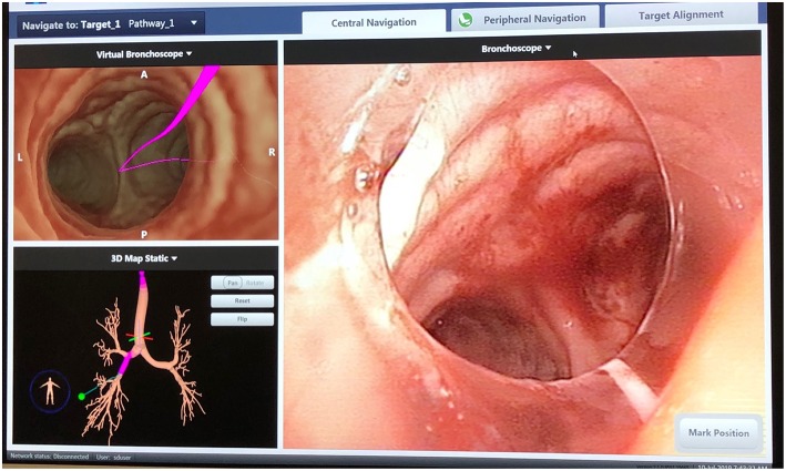
During minimally invasive pulmonary resection, it is often difficult to localize pulmonary nodules that are small (<2 cm), low-density/subsolid on imaging, or deep to the visceral pleura. The use of near-infrared fluorescence (NIF) imaging for localizing pulmonary nodules using indocyanine green (ICG) contrast is an emerging technology that is increasingly utilized during pulmonary resection. When administered via electromagnetic navigational bronchoscopy (ENB), ICG can accurately localize pulmonary nodules. When injected intravenously (IV), ICG can also help delineate the intersegmental plane. Research is ongoing regarding the utility of ICG for identification of the sentinel lymph node in lung cancer.

Keywords: electromagnetic; fluorescence; localization; lung cancer; navigational bronchoscopy; pulmonary resection; robotic.


Figures

Figure 1
Figure 1
SuperDimension Thoracic Navigation System: electromagnetic bronchoscopic view, showing the pathway to the target nodule in the right lower lobe.
Figure 2
Figure 2
SuperDimension Thoracic Navigation System: image-guided peripheral view, showing electromagnetic probe is 2 cm from the target nodule in the right lower lobe.
Figure 3
Figure 3
Pulmonary nodule in the left upper lobe, illuminated by ICG fluorescence (firefly mode).
Figure 4
Figure 4
Intravenous ICG delineation of the intersegmental plane between the lingula and the left upper lobe segments during left upper lobe trisegmentectomy. Note the diffusion of ICG contrast around the area of target nodule from Figure 3.

References

    1. Alander JT, Kaartinen I, Laakso A, Patila T, Spillmann T, Tuchin VV, et al. . A review of indocyanine green fluorescent imaging in surgery. Int J Biomed Imag. (2012) 2012:940585. 10.1155/2012/940585 - DOI - PMC - PubMed
    1. Aberle DR, Adams AM, Berg CD, Black WC, Clapp JD, Fagerstrom RM, et al. . Reduced lung-cancer mortality with low-dose computed tomographic screening. N Engl J Med. (2011) 365:395–409. 10.1056/NEJMoa1102873 - DOI - PMC - PubMed
    1. Ginsberg RJ, Rubinstein LV. Randomized trial of lobectomy versus limited resection for T1 N0 non-small cell lung cancer. Lung Cancer Study Group. Ann Thorac Surg. (1995) 60:615–22; discussion: 22–3. 10.1016/0003-4975(95)00537-U - DOI - PubMed
    1. Kodama K, Higashiyama M, Okami J, Tokunaga T, Imamura F, Nakayama T, et al. . Oncologic outcomes of segmentectomy versus lobectomy for clinical T1a N0 M0 Non-small cell lung cancer. Ann Thorac Surg. (2016) 101:504–11. 10.1016/j.athoracsur.2015.08.063 - DOI - PubMed
    1. Asamura H. Role of limited sublobar resection for early-stage lung cancer: steady progress. J Clin Oncol. (2014) 32:2403–4. 10.1200/JCO.2014.56.4203 - DOI - PubMed

LinkOut - more resources