. 2019 Sep 18;11(37):33620-33627.
doi: 10.1021/acsami.9b09983. Epub 2019 Sep 6.

Artificial Cell Membranes Interfaced with Optical Tweezers: A Versatile Microfluidics Platform for Nanomanipulation and Mechanical Characterization

Affiliations

Artificial Cell Membranes Interfaced with Optical Tweezers: A Versatile Microfluidics Platform for Nanomanipulation and Mechanical Characterization

Aurora Dols-Perez et al. ACS Appl Mater Interfaces. .

Abstract

Cell lipid membranes are the site of vital biological processes, such as motility, trafficking, and sensing, many of which involve mechanical forces. Elucidating the interplay between such bioprocesses and mechanical forces requires the use of tools that apply and measure piconewton-level forces, e.g., optical tweezers. Here, we introduce the combination of optical tweezers with free-standing lipid bilayers, which are fully accessible on both sides of the membrane. In the vicinity of the lipid bilayer, optical trapping would normally be impossible due to optical distortions caused by pockets of the solvent trapped within the membrane. We solve this by drastically reducing the size of these pockets via tuning of the solvent and flow cell material. In the resulting flow cells, lipid nanotubes are straightforwardly pushed or pulled and reach lengths above half a millimeter. Moreover, the controlled pushing of a lipid nanotube with an optically trapped bead provides an accurate and direct measurement of important mechanical properties. In particular, we measure the membrane tension of a free-standing membrane composed of a mixture of dioleoylphosphatidylcholine (DOPC) and dipalmitoylphosphatidylcholine (DPPC) to be 4.6 × 10⁻⁶ N/m. We demonstrate the potential of the platform for biophysical studies by inserting the cell-penetrating trans-activator of transcription (TAT) peptide in the lipid membrane. The interactions between the TAT peptide and the membrane are found to decrease the value of the membrane tension to 2.1 × 10⁻⁶ N/m. This method is also fully compatible with electrophysiological measurements and presents new possibilities for the study of membrane mechanics and the creation of artificial lipid tube networks of great importance in intra- and intercellular communication.

Keywords: cell membrane; lipid bilayer; lipid nanotube; microdevice; surface tension.


Conflict of interest statement

The authors declare no competing financial interest.

Figures

Figure 1
Figure 1
Design of the microdevice combining free-standing membranes with optical tweezers. (a) Picture of a representative microfluidic device used for mechanical measurements. Free-standing lipid bilayers are formed over the apertures connecting the two microchannels. The white square indicates the position of one of the apertures. (b, c) Pictures of the process of membrane formation (b) before the organic solvent reaches the aperture and (c) after membrane formation (A, air; O, organic phase; and W, aqueous phase; white arrows indicate the direction of the flow).
Figure 2
Figure 2
Effect of the lipid membrane annulus on optical imaging and optical trap stiffness. (a, b) Bright-field (left) and confocal fluorescence optical microscopy (right) images of lipid membranes formed using (a) a mixture of decane/chloroform/methanol as an organic solvent and (b) only chloroform as an organic solvent. (c, d) Pictures of a trapped bead near a membrane prepared using (c) decane/chloroform/methanol mixture and (d) chloroform. The distances between the bead and the membrane are indicated at the bottom of the pictures. (e) Optical trap stiffness in the x-axis, perpendicular to the membrane plane, as a function of the trap position with respect to the membrane. Trap stiffness measurements are all done with 1 μm beads and a laser power of 1.3 W (measured before entering the microscope objective) near the membranes formed using decane/chloroform/methanol (red circles) and chloroform (blue squares). The position represents the distance between the trap center and the membrane. The bars represent the standard deviation between measurements.
Figure 3
Figure 3
Lipid tube formation. (a) Bright-field images of a lipid tube formed by pulling a patch of membrane with an optically-trapped bead. The bead is first moved toward the membrane and then pulled away, as shown by the blue arrows. (b) Bright-field images of a lipid tube formed by pushing a bead against a free-standing lipid bilayer. (c) Bright-field images of two separate lipid tubes held by two optical traps. From top to bottom, the traps are brought closer to one another, as shown with blue arrows, until the two tubes contact and coalesce. (d) Six representative force–displacement curves obtained when pushing a 2 μm bead against the same lipid membrane.
Figure 4
Figure 4
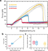
Force measurements when pushing beads of various sizes against a DOPC/DPPC lipid bilayer. (a) Force–displacement curves for tubes formed by pushing beads of 1, 2, and 5 μm diameters (N = 10, 15, and 14 curves, respectively), with representative curves shown in red, blue, and yellow, respectively, and all other curves shown in gray. (b) Maximum force and (c) tube extension force as a function of bead diameter.
Figure 5
Figure 5
Membrane properties are extracted from force curves. (a) Representative image from video recordings used to measure the angle θ of the membrane at a radial distance δ from the center of the bead. The shown force balance is used to measure the membrane tension. (b) Membrane tension, (c) bending rigidity, and (d) radius of lipid nanotubes are not statistically different for bead diameters 1, 2, and 5 μm (Kruskal–Wallis one-way analysis of variance, p > 0.05).
Figure 6
Figure 6
Effect of TAT peptide on membrane properties. (a) Force–displacement curves for tubes formed by pushing beads of 2 μm diameter against a DOPC/DPPC lipid bilayer without (gray) and with (green) TAT peptides, N = 15 and 16 curves, respectively. (b) Membrane tension extracted from the force–displacement curves.

Similar articles

Cited by

References

    1. Thottacherry J. J.; Kosmalska A. J.; Kumar A.; Vishen A. S.; Elosegui-Artola A.; Pradhan S.; Sharma S.; Singh P. P.; Guadamillas M. C.; Chaudhary N.; Vishwakarma R.; Trepat X.; del Pozo M. A.; Parton R. G.; Rao M.; Pullarkat P.; Roca-Cusachs P.; Mayor S. Mechanochemical Feedback Control of Dynamin Independent Endocytosis Modulates Membrane Tension in Adherent Cells. Nat. Commun. 2018, 9, 421710.1038/s41467-018-06738-5. - DOI - PMC - PubMed
    1. Gauthier N. C.; Fardin M. A.; Roca-Cusachs P.; Sheetz M. P. Temporary Increase in Plasma Membrane Tension Coordinates the Activation of Exocytosis and Contraction During Cell Spreading. Proc. Nat. Acad. Sci. U.S.A. 2011, 108, 14467–14472. 10.1073/pnas.1105845108. - DOI - PMC - PubMed
    1. Apodaca G. Modulation of Membrane Traffic by Mechanical Stimuli. Am. J. Physiol. Renal Physiol. 2002, 282, F179–F190. 10.1152/ajprenal.2002.282.2.F179. - DOI - PubMed
    1. Wang N.; Tytell J. D.; Ingber D. E. Mechanotransduction at a Distance: Mechanically Coupling the Extracellular Matrix with the Nucleus. Nat. Rev. Mol. Cell Biol. 2009, 10, 75–82. 10.1038/nrm2594. - DOI - PubMed
    1. Raucher D.; Sheetz M. P. Membrane Expansion Increases Endocytosis Rate During Mitosis. J. Cell. Biol. 1999, 144, 497–506. 10.1083/jcb.144.3.497. - DOI - PMC - PubMed

MeSH terms

Substances