

doi: 10.3791/59836.
Pan-myeloid Differentiation of Human Cord Blood Derived CD34+ Hematopoietic Stem and Progenitor Cells
Affiliations
- PMID: 31449258
- PMCID: PMC7046082
- DOI: 10.3791/59836
Item in Clipboard
Pan-myeloid Differentiation of Human Cord Blood Derived CD34+ Hematopoietic Stem and Progenitor Cells
J Vis Exp.
.
Abstract
Ex vivo differentiation of human hematopoietic stem cells is a widely used model for studying hematopoiesis. The protocol described here is for cytokine induced differentiation of CD34+ hematopoietic stem and progenitor cells to the four myeloid lineage cells. CD34+ cells are isolated from human umbilical cord blood and co-cultured with MS-5 stromal cells in the presence of cytokines. Immunophenotypic characterization of the stem and progenitor cells, and the differentiated myeloid lineage cells are described. Using this protocol, CD34+ cells may be incubated with small molecules or transduced with lentiviruses to express myeloid disease mutations to investigate their impact on myeloid differentiation.
Figures
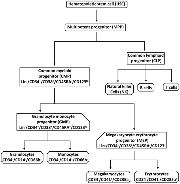
The pluripotent hematopoietic stem cell (HSC) differentiates into the multipotent progenitor (MPP), which gives rise to the common myeloid progenitor (CMP) and the common lymphoid progenitor (CLP) cells. CMPs generate two other myeloid progenitors, the granulocyte monocyte progenitors (GMPs) and the megakaryocyte erythrocyte progenitors (MEPs). Granulocytes and monocytes arise from GMPs, and erythrocytes and megakaryocytes arise from MEPs. CLPs give rise to natural killer, B, and T cells. The cell surface markers used to characterize the cell populations in this protocol are indicated. This figure has been modified from Bapat et al..
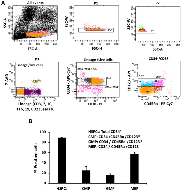
(A) Cells were gated on forward and side scatter to select a single cell population. Dead and lineage positive cells were eliminated by staining with 7-AAD and antibodies to CD3, CD7, CD10, CD11b, CD19, and CD235a (all FITC stained). The Lin−/live cells were analyzed with antibodies to CD34 (APC-Cy7), CD38 (PE), CD123 (APC), and CD45RA (PE-Cy7). The progenitors were distinguished from the CD34+/CD38+ cells. CMPs are CD123lo/CD45RA−, GMPs are CD123lo/CD45RA+ and MEPs are CD123−/CD45RA−. Representative scatter plots from an experiment are shown. (B) Percentages of total HSPCs (total CD34+ cells), CMPs, GMPs, and MEPs in cord blood are represented. Data presented are averages with standard error from at least 3 experiments.
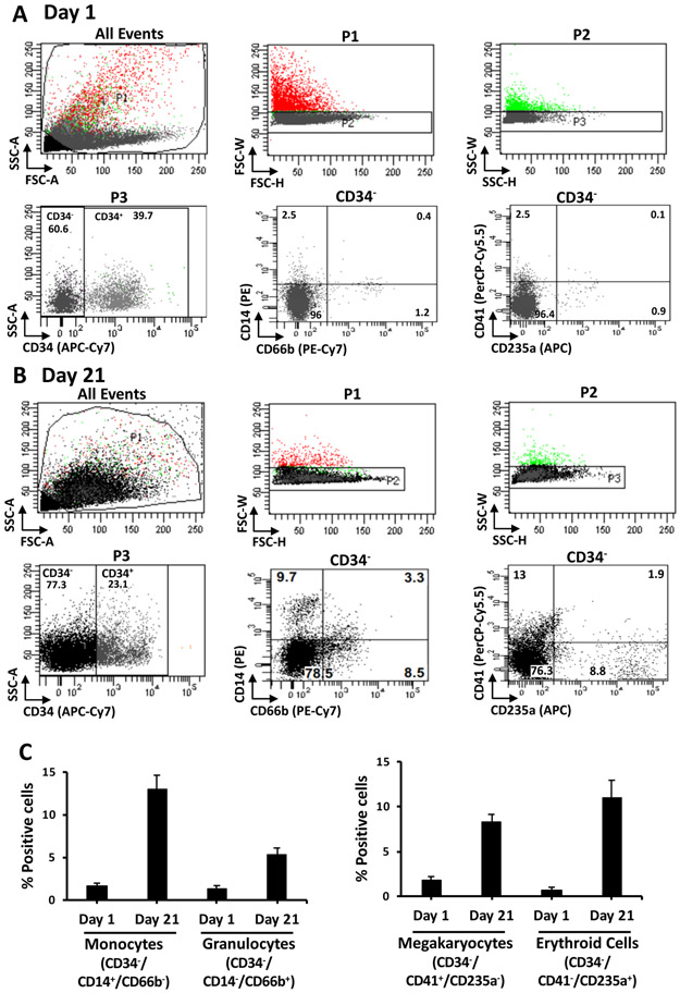
Single cells were gated based on forward and side scatter. CD34− cells were selected by staining with antibody to CD34 (APC-Cy7). Myeloid lineages in the CD34− population were analyzed with antibodies to CD14 (PE), CD66b (PE-Cy7), CD41 (PerCP-Cy5.5), and CD235a (APC) on day 1 (A) and day 21 (B). Monocytes are CD14+/CD66b−, granulocytes are CD14−/CD66b+, megakaryocytes are CD41+/CD235a− and erythroid cells are CD41−/CD235a+. Representative scatter plots from an experiment are shown. Fractions of monocytes, granulocytes, megakaryocytes, and erythroid cells in the CD34− population on day 1 and day 21 are represented (C). Data presented are averages with standard error from at least 3 experiments. This figure has been modified from Bapat et al..
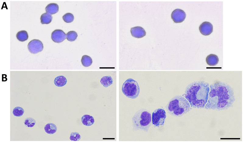
CD34+ HSPCs (A) and cells from myeloid cultures at day 21 (B) were stained with the Wright-Giemsa stain. Cells corresponding to granulocytes (black arrows), monocytes (blue arrows) and erythroid cells (red arrows) are shown. Scale bars represent 10 μM. This figure has been modified from Bapat et al..
References
-
- Hao QL, Shah AJ, Thiemann FT, Smogorzewska EM & Crooks GM A functional comparison of CD34 + CD38- cells in cord blood and bone marrow. Blood. 86 (10), 3745–3753, (1995). - PubMed
-
- Kondo M, Weissman IL & Akashi K Identification of clonogenic common lymphoid progenitors in mouse bone marrow. Cell. 91 (5), 661–672, (1997). - PubMed
-
- Haas S et al. Inflammation-Induced Emergency Megakaryopoiesis Driven by Hematopoietic Stem Cell-like Megakaryocyte Progenitors. Cell Stem Cell. 17 (4), 422–434, (2015). - PubMed
Publication types
MeSH terms
Substances
Grants and funding
LinkOut - more resources
Full Text Sources
Other Literature Sources
Medical
