. 2019 Oct 22;13(10):11273-11282.
doi: 10.1021/acsnano.9b04384. Epub 2019 Aug 26.

Needle-Free Injection of Exosomes Derived from Human Dermal Fibroblast Spheroids Ameliorates Skin Photoaging

Affiliations

Needle-Free Injection of Exosomes Derived from Human Dermal Fibroblast Spheroids Ameliorates Skin Photoaging

Shiqi Hu et al. ACS Nano. .

Abstract

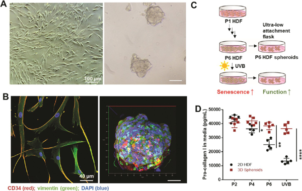
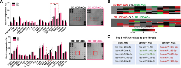
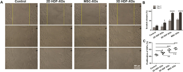
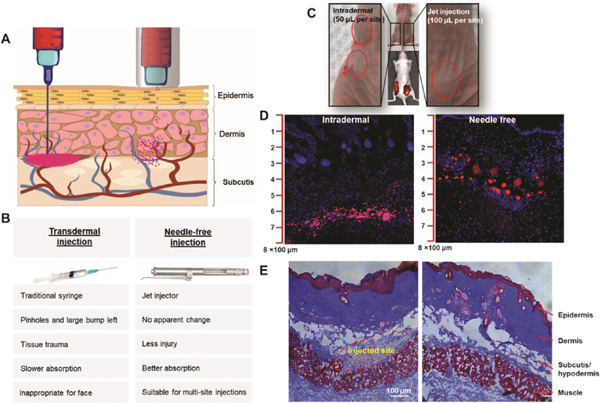
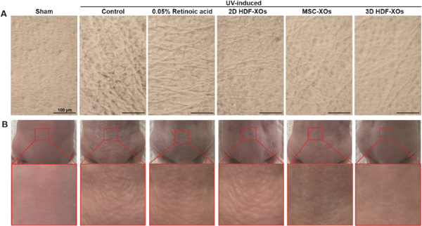
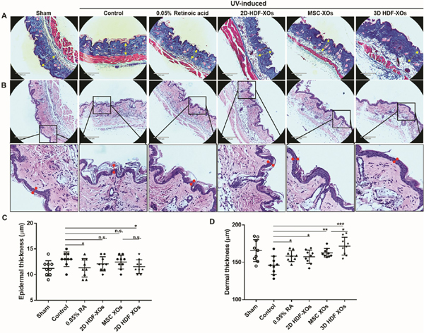
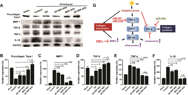
Human dermal fibroblasts (HDFs), the main cell population of the dermis, gradually lose their ability to produce collagen and renew intercellular matrix with aging. One clinical application for the autologous trans-dermis injection of HDFs that has been approved by the Food and Drug Administration aims to refine facial contours and slow down skin aging. However, the autologous HDFs used vary in quality according to the state of patients and due to many passages they undergo during expansion. In this study, factors and exosomes derived from three-dimensional spheroids (3D HDF-XOs) and the monolayer culture of HDFs (2D HDF-XOs) were collected and compared. 3D HDF-XOs expressed a significantly higher level of tissue inhibitor of metalloproteinases-1 (TIMP-1) and differentially expressed miRNA cargos compared with 2D HDF-XOs. Next, the efficacy of 3D HDF-XOs in inducing collagen synthesis and antiaging was demonstrated in vitro and in a nude mouse photoaging model. A needle-free injector was used to administer exosome treatments. 3D HDF-XOs caused increased procollagen type I expression and a significant decrease in MMP-1 expression, mainly through the downregulation of tumor necrosis factor-alpha (TNF-α) and the upregulation of transforming growth factor beta (TGF-β). In addition, the 3D-HDF-XOs group showed a higher level of dermal collagen deposition than bone marrow mesenchymal stem cell-derived exosomes. These results indicate that exosomes from 3D cultured HDF spheroids have anti-skin-aging properties and the potential to prevent and treat cutaneous aging.

Keywords: dermal fibroblasts; exosomes; microRNA; needle-free injection; skin aging; spheroids.


Figures

Figure 1.
Figure 1.
Comparison of 2D HDFs and 3D spheroids. (A) Photographs of human dermal fibroblasts (HDFs) and formed spheroids (scale bar: 100 μm). (B) Evaluation of vimentin (green channel) and CD34 (red channel) expression in 2D and 3D cultured HDFs (P6). DAPI (blue) was used to locate the nuclei of the cells (scale bar: 40 μm). (C) Schematic illustration of the culture process. (D) Pro-collagen I expression in 2D HDFs and spheroids from passages 2, 4, and 6, and after UVB exposure. Expression assessed by ELISA. n = 5, *p < 0.05,**p < 0.01, ****p < 0.0001.
Figure 2.
Figure 2.
Comparison of exosomes derived from MSCs and 2D and 3D HDFs. (A) Cytokine array of 2D HDF-XOs and 3D HDF-XOs (P6) by densitometric analysis (n = 3). (B) Heatmap of a fibrosis-related miRNA array incubated with 2D HDF-XOs, 3D spheroids-XOs, and MSC-XOs (n = 3). In 3D HDF-XOs, hsa-miR-196a-5p and hsa-miR-744-5p were downregulated compared to 2D HDF-XOs, while hsa-miR133a-3p, hsa-miR-223-3p, hsa-5011-5p, hsa-miR-325, hsa-miR-199b-5p, and hsa-miR-34a-5p were upregulated compared to both MSC-XOs and 2D HDF-XOs. (C) miRNAs of 2D HDF-XOs, 3D HDF-XOs, and MSC-XOs expressed at relatively high levels. n = 3, *p < 0.05, **p < 0.01.
Figure 3.
Figure 3.
Effects of exosomes on HDFs. (A) Wound recovery rates of HDFs, modeled by cell scratch assays. (B) The scratch closure rate is presented over time (n = 3). (C) HDF proliferation with the treatment of different exosomes, n = 3, n.s. means no significant difference, *p < 0.05, **p < 0.01, ***p < 0.001, ****p < 0.0001.
Figure 4.
Figure 4.
Comparison of intradermal injection with a syringe to needle-free injection with a jet injector. (A) Schematic illustration of needle injection and needle-free injection. (B) Comparison of syringe and jet injector properties and applications. (C) Exosomes were labeled with DiD (1,1′-dioctadecyl-3,3,3′,3′-tetramethylindodicarbocyanine, 4-chlorobenzenesulfonate salt) and injected into the dorsal skin of a nude mouse. The intradermal injections were administered on the left side of the back. The arrows indicate the injection sites. The jet injections were administered on the right side of the back. There was no obvious administration injury. A relatively large amount of solution pools in the tissue as a result of needle injections, which leads to local tissue trauma. Jet injections result in wider penetration and better absorption. (D) The mice were sacrificed 12 h after injection. Skin slices from the left and right sites were imaged via confocal microscopy. Highly concentrated exosomes (red) accumulated between the dermis and hypodermis in the mice that were intradermally injected. In mice treated with the jet injector, the exosomes dispersed well in both the dermis and hypodermis. (E) Representative skin histology. Scale bar: 100 μm.
Figure 5.
Figure 5.
Effects of retinoic acid (RA) and exosomes from different cells on wrinkle formation in UVB-irradiated nude mice. (A) Microscopic observation of replicas (scale bar: 100 μm) and (B) photographs of dorsal skin of mice from different groups (n = 3).
Figure 6.
Figure 6.
Histological analysis of the dorsal surface of treated and untreated nude mice after UVB irradiation. (A) Masson’s Trichrome staining. From left to right: sham, saline, dermal application of 0.05% retinoic acid (every other day), PRP, MSC-XOs/PRP, and 3D spheroids XOs/PRP (last three received one-time injections); scale bar: 290 μm. (B) Corresponding H&E staining, scale bar: 290 μm. (C) Epidermal and (D) dermal thickness analysis. n = 9 (3 mice per group, 3 spots analyzed for each sample), n.s. means no significant difference, *p < 0.05, **p < 0.01, ***p < 0.001.
Figure 7.
Figure 7.
Antiphotoaging mechanism signaling pathway analysis. (A) Western blot of dorsal skin of different groups. (B–F) Quantification of procollagen 1, MMP1, TGF-β, TNF-α, and IL-1β (n = 3); n.s. means no significant difference, *p < 0.05, **p < 0.01, ***p < 0.001, ****p < 0.0001. (G) Schematic illustration of the mechanism of 3D HDF-XOs treatment.

References

    1. Castleberry SA; Quadir MA; Sharkh MA; Shopsowitz KE; Hammond PT Polymer Conjugated Retinoids for Controlled Transdermal Delivery. J. Controlled Release 2017, 262, 1–9. - PMC - PubMed
    1. Schagen S Topical Peptide Treatments with Effective Anti-Aging Results. Cosmetics 2017, 4, 16–29.
    1. Aldag C; Teixeira DN; Leventhal PS Skin Rejuvenation Using Cosmetic Products Containing Growth Factors, Cytokines, and Matrikines: a Review of the Literature. Clin., Cosmet. Invest. Dermatol. 2016, 9, 411–419. - PMC - PubMed
    1. Kim JH; Kwon T-R; Hong SW; Seok J; Kim JM; Hong JY; Lee SE; Han SW; Kim BJ Comparative Evaluation of the Biodegradability and Wrinkle Reduction Efficacy of Human-Derived Collagen Filler and Hyaluronic Acid Filler. Aesthetic Plast. Surg. 2019, 43, 1095–1101. - PubMed
    1. Jeong JH; Fan Y; You GY; Choi TH; Kim S Improvement of Photoaged Skin Wrinkles with Cultured Human Fibroblasts and Adipose-Derived Stem Cells: a Comparative Study. J. Plast. Reconstr. Aesthet. Surg. 2015, 68, 372–381. - PubMed

Publication types

MeSH terms

Substances