Review
. 2019 Aug 24;24(17):3074.
doi: 10.3390/molecules24173074.

Structure and Function of Multimeric G-Quadruplexes

Affiliations
Review

Structure and Function of Multimeric G-Quadruplexes

Sofia Kolesnikova et al. Molecules. .

Abstract

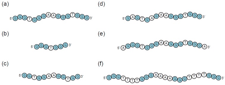
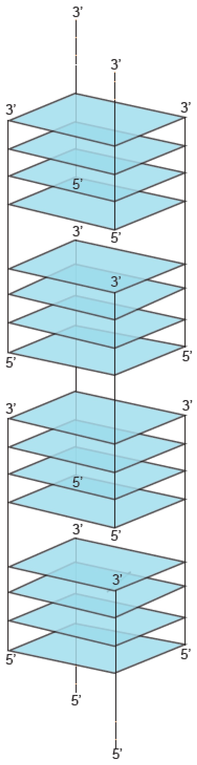
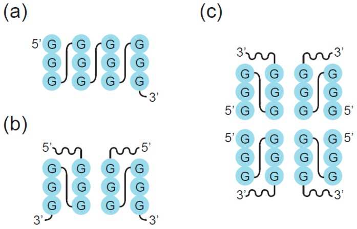
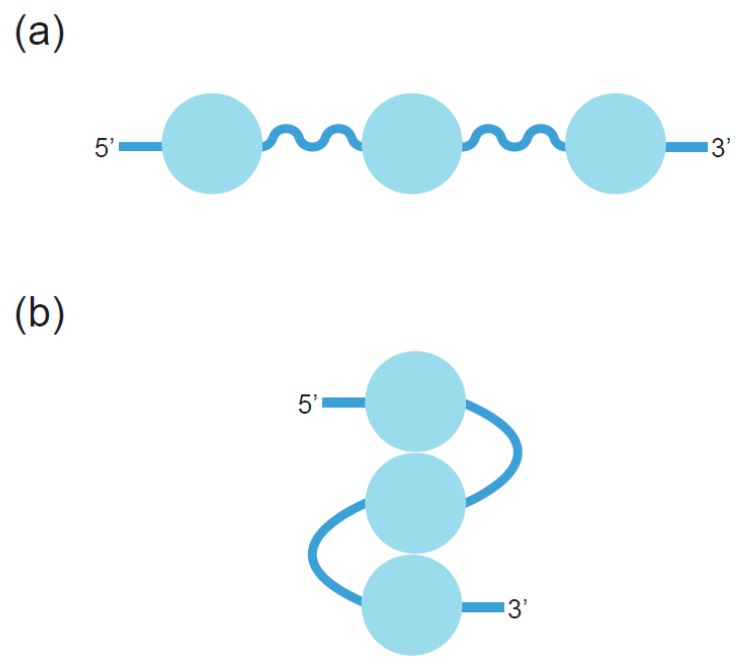
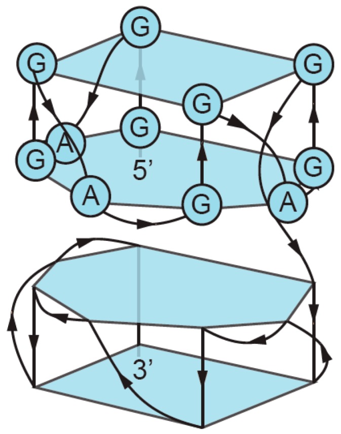
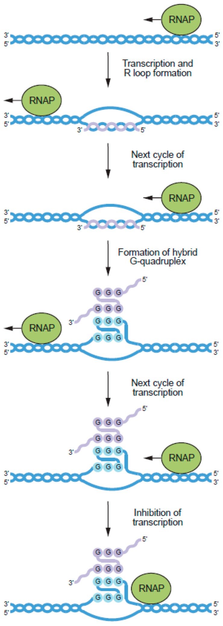
G-quadruplexes are noncanonical nucleic acid structures formed from stacked guanine tetrads. They are frequently used as building blocks and functional elements in fields such as synthetic biology and also thought to play widespread biological roles. G-quadruplexes are often studied as monomers, but can also form a variety of higher-order structures. This increases the structural and functional diversity of G-quadruplexes, and recent evidence suggests that it could also be biologically important. In this review, we describe the types of multimeric topologies adopted by G-quadruplexes and highlight what is known about their sequence requirements. We also summarize the limited information available about potential biological roles of multimeric G-quadruplexes and suggest new approaches that could facilitate future studies of these structures.

Keywords: DNA:RNA hybrid; G-quadruplex; R-loop; dimer; multimer; oligomer; promoter; telomere; tetramer.


Conflict of interest statement

The authors declare no conflict of interest.

Figures

Figure 1
Figure 1
Regulation of biochemical function by multimerization. (a) Concentration-based control of biochemical function. In this scheme, monomers are biochemically active and dimers are not. At concentrations below the dissociation constant for dimer formation, most of the population is monomeric and in the active state, while at concentrations above the dissociation constant, most of the population is dimeric and in the inactive state. Green rectangles = nucleic acid or protein monomers. (b) Enhanced sensitivity to ligand concentration by cooperative binding. In this scheme, ligand binding is independent when nucleic acid or protein binding sites are monomeric but cooperative when they are linked by multimerization. This leads to all-or-none binding and enhanced sensitivity to ligand concentration. Green rectangles = nucleic acid or protein monomers; black circles = ligands. (c) Modulation of biochemical activity by the exchange of dimerization partners. In this scheme, the gene transcribed by RNA polymerase is determined by the DNA-binding specificity of its dimerization partner. Green ovals = RNA polymerase; green and purple rectangles = transcription factors; blue lines = DNA.
Figure 2
Figure 2
Chemical structure of a GQ (G-quadruplex) tetrad.
Figure 3
Figure 3
Formation of GQs from different numbers of strands. (a) Intramolecular (unimolecular) GQ with antiparallel strands. (b) Intramolecular (unimolecular) GQ with parallel strands. (c) Bimolecular (dimeric) GQ. (d) Trimolecular (trimeric) GQ. (e) Tetramolecular (tetrameric) GQ. Note that each of these structures can in principle contain all parallel strands, all antiparallel strands, or a mix of parallel and antiparallel strands.
Figure 4
Figure 4
Types of interfaces in multimeric GQs. (a) First mode of multimerization. Interfaces are formed between tetrads which stack on top of one another in a 5′ to 5′, 3′ to 3′, or 5′ to 3′ arrangement. (b) Second mode of multimerization. Interfaces are formed within tetrads made up of guanines from multiple DNA strands. (c) Structure combining these two modes of multimerization.
Figure 5
Figure 5
Sequence requirements of multimeric GQs. (a) Example of a canonical GQ. (b) Variant containing two rather than four G-runs. Such a sequence can form a multimeric GQ but not a monomeric one. (c) Variant containing G-runs of two rather than three nucleotides. In some cases, such sequences form multimeric rather than monomeric structures. (d) Variant containing mutations in tetrads. Such mutations can induce formation of dimeric and tetrameric structures. (e) Variant containing overhanging nucleotides. Such variants typically cannot stack via the first mode of multimerization, but the ability to interact via the second mode of multimerization is unaffected. (f) Variant containing extended loops. Longer loops favor formation of antiparallel rather than parallel GQs, and such loops can interfere with the ability of GQs to stack via the first mode of multimerization.
Figure 6
Figure 6
Structure of a G-wire.
Figure 7
Figure 7
Mutations in tetrads can induce GQ multimerization. (a) Secondary structure of a GQ we are studying in our group with the sequence GGGTGGGAAGGGTGGGA. We previously generated a library containing all possible mutations in the central tetrad in this structure (at positions 2, 6, 11, and 15) and tested these variants for a series of biochemical activities associated with GQs [18,77,78,79]. (b) Proposed secondary structure of dimers formed by variants containing mutations at positions 2, 6, or both in the central tetrad of the reference GQ. The 5′ part of this structure, which contains the mutated nucleotides, is represented by a wavy black line. (c) Proposed secondary structure of tetramers formed by variants containing mutations at positions 11, 15, or both in the central tetrad of the reference GQ. The 3′ part of this structure, which contains the mutated nucleotides, is represented by a wavy black line.
Figure 8
Figure 8
Possible structures of telomeric GQs. (a) Beads-on-a-string model in which telomeric GQs do not interact. (b) Model in which telomeric GQs stack on one another to form higher-order structures. Blue circles represent GQ structures.
Figure 9
Figure 9
Structure of a dimeric GQ formed by the sequence (GGA)8. See [64] and [107] for more information about this structure.
Figure 10
Figure 10
Model for the regulation of transcription by DNA:RNA hybrid GQs, formed between the noncoding strands of G-rich genes and RNA molecules transcribed from these genes. The newly synthesized RNA transcript is shown in purple. See [130] for more information about this model.

References

    1. Rich A. DNA comes in many forms. Gene. 1993;135:99–109. doi: 10.1016/0378-1119(93)90054-7. - DOI - PubMed
    1. Hu Y., Cecconello A., Idili A., Ricci F., Willner I. Triplex DNA nanostructures: From basic properties to applications. Angew. Chem. Int. Ed. Engl. 2017;56:15210–15233. doi: 10.1002/anie.201701868. - DOI - PubMed
    1. Davis J.T. G-quartets 40 years later: From 5′-GMP to molecular biology and supramolecular chemistry. Angew. Chem. Int. Ed. Engl. 2004;43:668–698. doi: 10.1002/anie.200300589. - DOI - PubMed
    1. Day H.A., Pavlou P., Waller Z.A. I-Motif DNA: Structure, stability and targeting with ligands. Bioorg. Med. Chem. 2014;22:4407–4418. doi: 10.1016/j.bmc.2014.05.047. - DOI - PubMed
    1. Burge S., Parkinson G.N., Hazel P., Todd A.K., Neidle S. Quadruplex DNA: Sequence, topology and structure. Nucleic Acids Res. 2006;34:5402–5415. doi: 10.1093/nar/gkl655. - DOI - PMC - PubMed