Review
. 2019 Aug 24;10(9):640.
doi: 10.3390/genes10090640.

Effects of Radiation Therapy on Neural Stem Cells

Affiliations
Review

Effects of Radiation Therapy on Neural Stem Cells

Anna Michaelidesová et al. Genes (Basel). .

Abstract

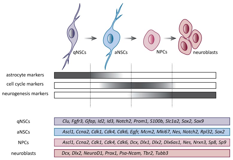
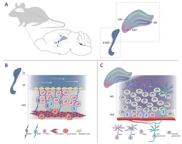
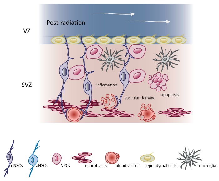
Brain and nervous system cancers in children represent the second most common neoplasia after leukemia. Radiotherapy plays a significant role in cancer treatment; however, the use of such therapy is not without devastating side effects. The impact of radiation-induced damage to the brain is multifactorial, but the damage to neural stem cell populations seems to play a key role. The brain contains pools of regenerative neural stem cells that reside in specialized neurogenic niches and can generate new neurons. In this review, we describe the advances in radiotherapy techniques that protect neural stem cell compartments, and subsequently limit and prevent the occurrence and development of side effects. We also summarize the current knowledge about neural stem cells and the molecular mechanisms underlying changes in neural stem cell niches after brain radiotherapy. Strategies used to minimize radiation-related damages, as well as new challenges in the treatment of brain tumors are also discussed.

Keywords: brain and nervous system cancers; neural stem cells; neurogenic niches; radiotherapy; sparing of neurogenic regions.


Conflict of interest statement

The authors declare no competing interests.

Figures

Figure 1
Figure 1
Cell subtypes involved in progression from quiescent neural stem cells (qNSCs) to neuroblasts. Schematic representation of lineage progression. QNSCs give rise to activated neural stem cells (aNSCs), which differentiate into highly proliferative progenitor cells (NPCs) and finally to neuroblasts. Expression of key genes related to particular cell subtypes is depicted. Ascl1, achaete-scute family bHLH transcription factor 1; Ccna2, cyclin A2; Cdk1, cyclin dependent kinase 1; Cdk4, cyclin dependent kinase 4; Cdk6, cyclin dependent kinase 6; Clu, clusterin; Dcx, doublecortin; Dlx1, distal-less homeobox 1; Dlx2, distal-less homeobox 2; Dlx6as1, distal-less homeobox 6, opposite strand 1; Egfr, epidermal growth factor receptor; Gfap, glial fibrillary acidic protein; Id2, inhibitor of DNA binding 2; Id3, inhibitor of DNA binding 3; Mcm2, minichromosome maintenance complex component 2; Mki67, antigen identified by monoclonal antibody Ki-67; Nes, nestin; NeuroD1, neurogenic differentiation 1; Notch2, notch 2; Nrxn3, neurexin 3; Prom1, prominin-1; Prox1, prospero homeobox 1; Psa-Ncam, polysialylated neural cell adhesion molecule; Rpl32, ribosomal protein L32; S100b, S100 protein, beta polypeptide, neural; Slc1a2, solute carrier family 1 (glial high affinity glutamate transporter), member 2; Sox2, SRY (sex determining region Y)-box 2; Sox9, SRY (sex determining region Y)-box 9; Sp8, trans-acting transcription factor 8; Sp9, trans-acting transcription factor 9; Tbr2, eomesodermin; Tubb3, tubulin, beta 3 class III.
Figure 2
Figure 2
Neurogenesis in adult mouse brain. (A) Sagittal view of adult mouse brain focusing on two neurogenic niches where NSCs reside—the ventricular-subventricular zone (V-SVZ) of the lateral ventricle (LV) and dentate gyrus (DG) of the hippocampus (H). Cornu Ammonis 1 (CA1) and Cornu Ammonis 3 (CA3) subfields of the hippocampus are depicted. (B) Schematic representation of the organization and composition of the adult mouse V-SVZ niche. qNSCs share many characteristics with aNSCs, including contact with blood vessels. White arrows show the flow of the cerebrospinal fluid. (C) Schematic representation of the cell types present in the mouse subgranular zone (SGZ) and granule cell layer (GCL) in the dentate gyrus of the hippocampus.
Figure 3
Figure 3
Radiation disrupts the V-SVZ niche. Schematic representation of mice V-SVZ niche after radiation. Following radiation, the V-SVZ niche shows a depletion of proliferating aNSCs, NPCs, and neuroblasts, a vascular damage and an increase in the number of microglia. Compare with schematic representation of mice V-SVZ niche in a pre-radiation condition (Figure 2B).

References

    1. Worldwide Cancer Statistics. [(accessed on 26 April 2019)]; Available online: https://www.cancerresearchuk.org/health-professional/cancer-statistics/w....
    1. Cancer Today. [(accessed on 26 April 2019)]; Available online: http://gco.iarc.fr/today/home.
    1. Behin A., Hoang-Xuan K., Carpentier A.F., Delattre J.Y. Primary brain tumours in adults. Lancet. 2003;361:323–331. doi: 10.1016/S0140-6736(03)12328-8. - DOI - PubMed
    1. Ostrom Q.T., Wright C.H., Barnholtz-Sloan J.S. Brain metastases: Epidemiology. Handb. Clin. Neurol. 2018;149:27–42. - PubMed
    1. Villano J.L., Durbin E.B., Normandeau C., Thakkar J.P., Moirangthem V., Davis F.G. Incidence of brain metastasis at initial presentation of lung cancer. Neuro-Oncology. 2015;17:122–128. doi: 10.1093/neuonc/nou099. - DOI - PMC - PubMed

Publication types