. 2019 Aug 24;8(9):971.
doi: 10.3390/cells8090971.

PI3K (Phosphatidylinositol 3-Kinase) Activation and Endothelial Cell Proliferation in Patients with Hemorrhagic Hereditary Telangiectasia Type 1

Affiliations

PI3K (Phosphatidylinositol 3-Kinase) Activation and Endothelial Cell Proliferation in Patients with Hemorrhagic Hereditary Telangiectasia Type 1

Adriana Iriarte et al. Cells. .

Abstract

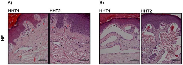
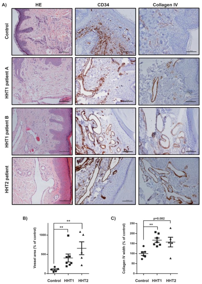
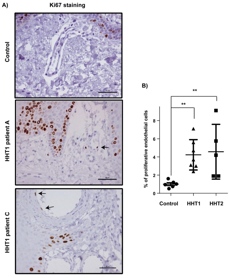
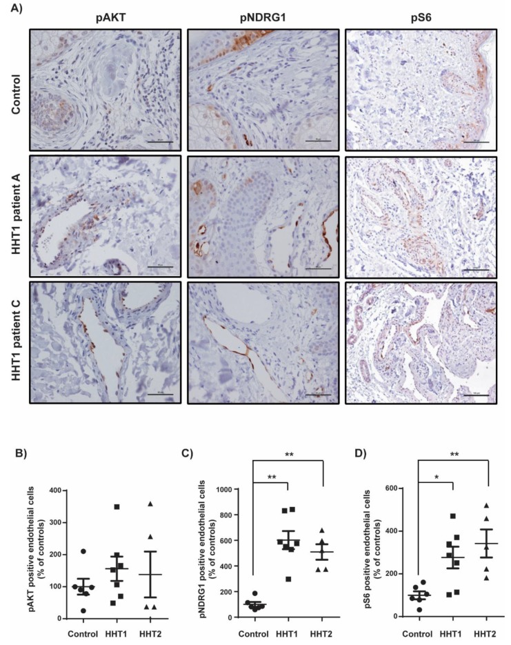
Hemorrhagic hereditary telangiectasia (HHT) type 2 patients have increased activation of the phosphatidylinositol 3-kinase (PI3K) signaling pathway in telangiectasia. The main objective is to evaluate the activation of the PI3K pathway in cutaneous telangiectasia of HHT1 patients. A cutaneous biopsy of a digital hand telangiectasia was performed in seven HHT1 and eight HHT2 patients and compared with six controls. The study was approved by the Clinical Research Ethics Committee of our center. A histopathological pattern with more dilated and superficial vessels that pushed up the epidermis was identified in HHT patients regardless of the type of mutation and was associated with older age, as opposed to the common telangiectasia pattern. The mean proliferation index (Ki-67) was statistically higher in endothelial cells (EC) from HHT1 than in controls. The percentage of positive EC for pNDRG1, pAKT, and pS6 in HHT1 patients versus controls resulted in higher values, statistically significant for pNDRG1 and pS6. In conclusion, we detected an increase in EC proliferation linked to overactivation of the PI3K pathway in cutaneous telangiectasia biopsies from HHT1 patients. Our results suggest that PI3K inhibitors could be used as novel therapeutic agents for HHT.

Keywords: Smad pathway; hemorrhagic hereditary telangiectasia; mTOR inhibitors; phosphatidylinositol 3-kinase; rare diseases; telangiectasia; transforming growth factor-beta (TGF-β).


Conflict of interest statement

The authors declare no conflict of interest.

Figures

Figure 1
Figure 1
Hematoxylin and eosin (HE) microscope images. Scale bars, 100 µm. (A) Examples of conventional telangiectasia characterized by the presence of dilated microvessels at the superficial dermis (black arrows). Normal vessels are indicated with red arrows. One HHT1 patient and one HHT2. (B) Examples of more dilatated vessels expanding the papillary dermis between rete ridges that crowd up the epidermis layer (black arrows) (angiokeratoma-like pattern). One HHT1 patient and one HHT2.
Figure 2
Figure 2
Increased vessels size and collagen IV staining in HHT1 and HHT2 cutaneous telangiectasia biopsies. (A) Hematoxylin and eosin (HE), CD34, and collagen IV staining of one control, two HHT1 patients and one HHT2 patient biopsies. Scale bars, 100 µm. (B) Quantification of the vessel area in controls (n = 6), HHT1 (n = 7), and HHT2 patients (n = 5). Results are represented as % relative to the control. Error bars indicate the standard error of the mean. Statistical significance of two-tailed Mann–Whitney U tests: ** p < 0.01. (C) Quantification of the collagen IV width in controls (n = 6), HHT1 (n = 7), and HHT2 patients (n = 5). Results are represented as % relative to the control. Error bars indicate the standard error of the mean. Statistical significance of two-tailed Mann–Whitney U tests: ** p < 0.01.
Figure 3
Figure 3
Increased endothelial cell proliferation in HHT1 cutaneous telangiectasia biopsies. (A) Ki-67 (brown nuclei, arrows) staining of endothelial cells in one control and two HHT1 patients. Scale bars, 100 µm. (B) Quantification of the percentage of Ki-67-positive endothelial cells in controls (n = 6), HHT1 patients (n = 7), and HHT2 patients (n = 5). Error bars indicate the standard errors of the mean. Statistical significance of two-tailed Mann–Whitney U tests: ** p < 0.01.
Figure 4
Figure 4
Increased activation of PI3K signaling in HHT1 cutaneous telangiectasia biopsies. (A) pAKT, pNDRG1, and pS6 staining of a control and two HHT1 patient biopsies. Scale bars, 100 µm. (B) Quantification of the percentage of pAKT-positive endothelial cells in controls (n = 6), HHT1 patients (n = 7), and HHT2 patients (n = 4). Results are represented as % relative to the control. Error bars indicate the standard error of the mean. (C) Quantification of the percentage of pNDRG1-positive endothelial cells in controls (n = 6), HHT1 patients (n = 7), and HHT2 patients (n = 5). Results are represented as % relative to the control. Error bars indicate the standard error of the mean. Statistical significance of two-tailed Mann–Whitney U tests: ** p < 0.01. (D) Quantification of the percentage of pS6-positive endothelial cells in controls (n = 6), HHT1 patients (n = 7), and HHT2 patients (n = 5). Results are represented as % relative to the control. Error bars indicate the standard error of the mean. Statistical significance of two-tailed Mann–Whitney U tests: * p < 0.05; ** p < 0.01.

References

    1. The Portal for Rare Diseases and Orphan Drugs. [(accessed on 22 July 2019)]; Available online: https://www.orpha.net/consor/cgi-bin/index.php.
    1. Abdalla S.A., Letarte M. Hereditary haemorrhagic telangiectasia: Current views on genetics and mechanisms of disease. J. Med. Genet. 2006;43:97–110. doi: 10.1136/jmg.2005.030833. - DOI - PMC - PubMed
    1. McDonald J., Wooderchak-Donahue W., VanSant Webb C., Whitehead K., Stevenson D.A., Bayrak-Toydemir P. Hereditary hemorrhagic telangiectasia: Genetics and molecular diagnostics in a new era. Front. Genet. 2015;6 doi: 10.3389/fgene.2015.00001. - DOI - PMC - PubMed
    1. Tillet E., Bailly S. Emerging roles of BMP9 and BMP10 in hereditary hemorrhagic telangiectasia. Front. Genet. 2015;5 doi: 10.3389/fgene.2014.00456. - DOI - PMC - PubMed
    1. Mahmoud M., Allinson K.R., Zhai Z., Oakenfull R., Ghandi P., Adams R.H., Fruttiger M., Arthur H.M. Pathogenesis of arteriovenous malformations in the absence of endoglin. Circ. Res. 2010;106:1425–1433. doi: 10.1161/CIRCRESAHA.109.211037. - DOI - PubMed

Publication types

MeSH terms

Substances

Supplementary concepts