Review
. 2019 Aug 25;20(17):4142.
doi: 10.3390/ijms20174142.

Molecular-Level Interactions between Engineered Materials and Cells

Affiliations
Review

Molecular-Level Interactions between Engineered Materials and Cells

Yoon-Ha Jang et al. Int J Mol Sci. .

Abstract

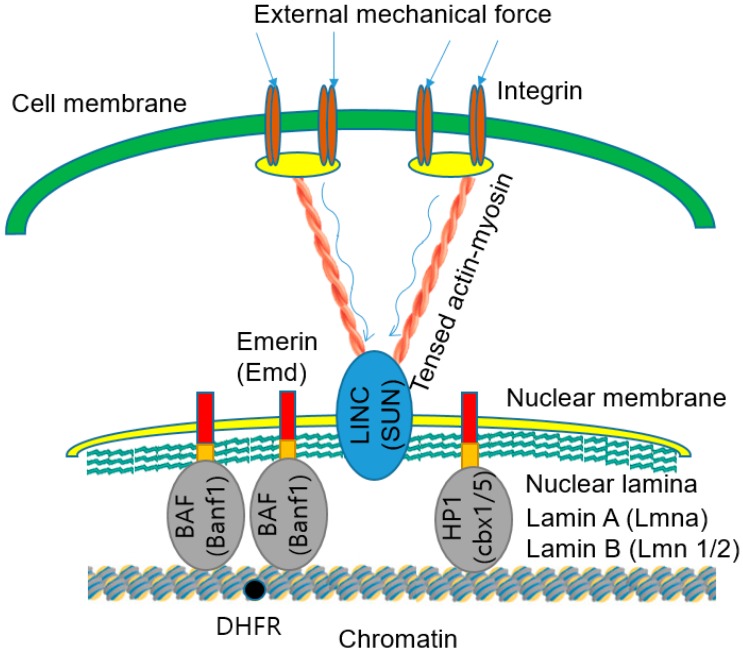
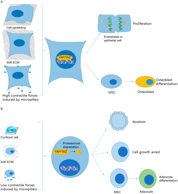
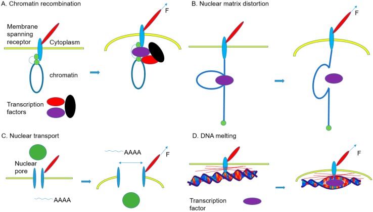
Various recent experimental observations indicate that growing cells on engineered materials can alter their physiology, function, and fate. This finding suggests that better molecular-level understanding of the interactions between cells and materials may guide the design and construction of sophisticated artificial substrates, potentially enabling control of cells for use in various biomedical applications. In this review, we introduce recent research results that shed light on molecular events and mechanisms involved in the interactions between cells and materials. We discuss the development of materials with distinct physical, chemical, and biological features, cellular sensing of the engineered materials, transfer of the sensing information to the cell nucleus, subsequent changes in physical and chemical states of genomic DNA, and finally the resulting cellular behavior changes. Ongoing efforts to advance materials engineering and the cell-material interface will eventually expand the cell-based applications in therapies and tissue regenerations.

Keywords: cell surface sensors; cellular responses; genome states; materials engineering; mechanotransduction.


Conflict of interest statement

The authors declare no conflict of interest.

Figures

Figure 1
Figure 1
Mo3Se3 SCAC-based extracellular matrix (ECM) mimicking materials [34]. (A) Properties of Mo3Se3 SCACs to mimic ECM. (B) ECM-mimicking scaffold prepared by spray coating of Mo3Se3 SCACs onto a glass substrate. (C) Proliferation of MC3T3-E1 cell lines treated with media containing different concentrations of Mo3Se3 SCACs (* for p < 0.0001). All statistical analysis was performed with the control data. “Reprinted with permission from ref. [34]. Copyright 2018, American Chemical Society.”
Figure 2
Figure 2
The effects of substrate geometry on cells. (A) The patterns of forces exerted by the cells responding to the edges and boundaries of different substrates. (a) Colorimetric stacked images of cell proliferation in a small (250 µm edge) square, (b) large (500 µm edge) square, (c) small (125 × 500 µm) rectangular, and (d) large (564 µm diameter) circular islands [37]. “Reprinted with permission from ref. [37]. Copyright 2005, National Academy of Sciences” (B) A model of geometrical, biochemical, and mechanical maturation of integrin-mediated cell adhesion and behaviour after responding to nanopatterned matrices [42]. “Reprinted with permission from [42]. Copyright 2014, American Chemical Society.” (C) Schematic representation of (a) the cytoskeletal forces acting on the nucleus (F-actin in red and lamin-A in green) and (b) the proposed geometry-induced changes in cellular attachment and forces on the nucleus for flat, concave and convex surfaces [43]. “Reprinted with permission from ref. [43]. Reproduced with permission under Creative Commons Attribution 4.0 International License http://creativecommons.org/licenses/by/4.0/.
Figure 3
Figure 3
Figure 3. Myotube alignment and orientation on engineered substrates. (A) The effects of substrate-bound adhesion molecules on alignment and orientation of myotubes. Eight hundred nanometer grooved substrates were functionalized with different ECM components. (B) Myotube alignment and orientation on 800 nm grooved Matrigel-functionalized substrates to differentiate diseased and nondiseased cells (inset: bright-field images show nanogroove directions (marked with red arrows)) [65]. “Reprinted from ref. [65], Copyright 2018, with permission from Elsevier.”
Figure 4
Figure 4
Conformational rearrangement of integrin upon attachment to substrate [79,80,81]. Integrin receptor proteins work as αβ heterodimers. When attaching to substrate, integrins undergo conformational rearrangements from bent conformation with low affinity for ligand to extended conformation with high affinity for ligand. This integrin binding to substrates induces signal transduction and provides physical connections between extracellular and intracellular regions through focal adhesion complex formation.
Figure 5
Figure 5
Propagation of external mechanical force from integrins to chromatin [124]. External mechanical forces applied to the cell surface are propagated via integrins and tensed actin-myosin cytoskeleton to LINC complexes and nuclear lamins in the nuclear lamina. The forces are then transferred to the chromatin through heterochromatin protein (HP1), Barrier-to-autointegration factor (BAF) proteins, and other molecules. The forces transferred to the chromatin stretch deform the chromatin segment that contains the DHFR gene. The deformation and stretching of chromatin facilitate binding of transcription factors to the DHFR gene for the upregulation of the DHFR gene transcription. The external mechanical force was provided by ferromagnetic beads attached to integrins in a magnetic field. The magnetic stress was 17.5 Pa.
Figure 6
Figure 6
Mechanical stimuli induce YAP/TAZ translocation into the nucleus and determine cell fates [113,147]. (A) The translocation of the transcriptional regulators, YAP and TAZ, into the nucleus occurs under mechanical conditions that induce strong intracellular resistive forces and activate YAP and TAZ. In cells spread on an extensive adhesive area, cultured on solid extracellular matrices (ECMs), or stretched between micropillars, YAP and TAZ translocate into the nucleus and become active. Under these conditions, these transcriptional regulators are required for endothelial or epithelial cell proliferation and differentiation of MSCs to osteoblasts. (B) The inactivation and relocalization of YAP and TAZ in cytoplasm followed by proteasomal degradation of YAP and TAZ occur when the cells are confined on small adhesive areas or cultured on soft ECMs or on top of micropillars. The degradation results in weak contractile forces. Degradation of YAP and TAZ causes cell apoptosis, growth arrest, or differentiation of MSCs into adipocytes. Furthermore, the degradation and nuclear localization of YAP and TAZ are affected by ECM properties such as stiffness, area, and contractile force.
Figure 7
Figure 7
Mechanisms of gene regulation by mechanical stimuli [153]. (A) External mechanical forces deform nuclear scaffolds and chromatin organization, thereby altering assembly of transcription factors for gene regulation. (B) Forces transferred to specific chromatin regions tethered to lamins or other nuclear membrane receptors regulate the activities of assembled transcription factors or splicing factors. (C) Forces applied to nuclear pores change the pore size, thereby altering nuclear transport and gene expression by influencing mRNA transport. (D) Forces transferred to DNA through nuclear scaffolds separate the DNA double helix at specific regions, facilitating the binding of transcription factors to the region.

References

    1. Park I.H., Lerou P.H., Zhao R., Huo H., Daley G.Q. Generation of human-induced pluripotent stem cells. Nat. Protoc. 2008;3:1180–1186. doi: 10.1038/nprot.2008.92. - DOI - PubMed
    1. Liu Y., Conaway L., Rutherford Bethard J., Al-Ayoubi A.M., Thompson Bradley A., Zheng H., Weed S.A., Eblen S.T. Phosphorylation of the alternative mRNA splicing factor 45 (SPF45) by Clk1 regulates its splice site utilization, cell migration and invasion. Nucleic Acids Res. 2013;41:4949–4962. doi: 10.1093/nar/gkt170. - DOI - PMC - PubMed
    1. Fu X., Zhang H., Chen Z., Yang Z., Shi D., Liu T., Chen W., Yao F., Su X., Deng W., et al. TFAP2B overexpression contributes to tumor growth and progression of thyroid cancer through the COX-2 signaling pathway. Cell Death Dis. 2019;10:397. doi: 10.1038/s41419-019-1600-7. - DOI - PMC - PubMed
    1. Cullot G., Boutin J., Toutain J., Prat F., Pennamen P., Rooryck C., Teichmann M., Rousseau E., Lamrissi-Garcia I., Guyonnet-Duperat V., et al. CRISPR-Cas9 genome editing induces megabase-scale chromosomal truncations. Nat. Commun. 2019;10:1136. doi: 10.1038/s41467-019-09006-2. - DOI - PMC - PubMed
    1. Yao C.H., Liu G.Y., Wang R., Moon S.H., Gross R.W., Patti G.J. Identifying off-target effects of etomoxir reveals that carnitine palmitoyltransferase I is essential for cancer cell proliferation independent of beta-oxidation. PLoS Biol. 2018;16:e2003782. doi: 10.1371/journal.pbio.2003782. - DOI - PMC - PubMed

Substances

LinkOut - more resources