. 2019 Aug 23;7(3):96.
doi: 10.3390/vaccines7030096.

Immunogenicity and Protection Efficacy of a Naked Self-Replicating mRNA-Based Zika Virus Vaccine

Affiliations

Immunogenicity and Protection Efficacy of a Naked Self-Replicating mRNA-Based Zika Virus Vaccine

Zifu Zhong et al. Vaccines (Basel). .

Abstract

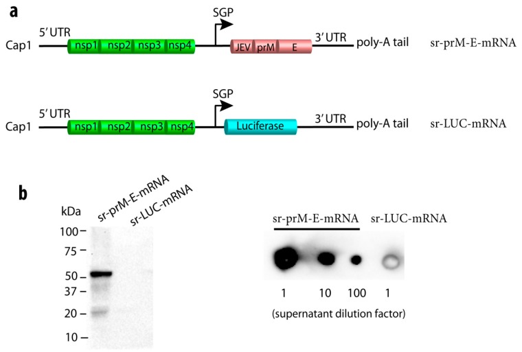
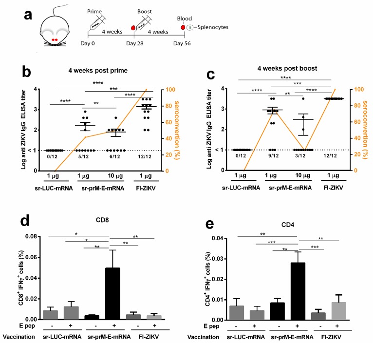
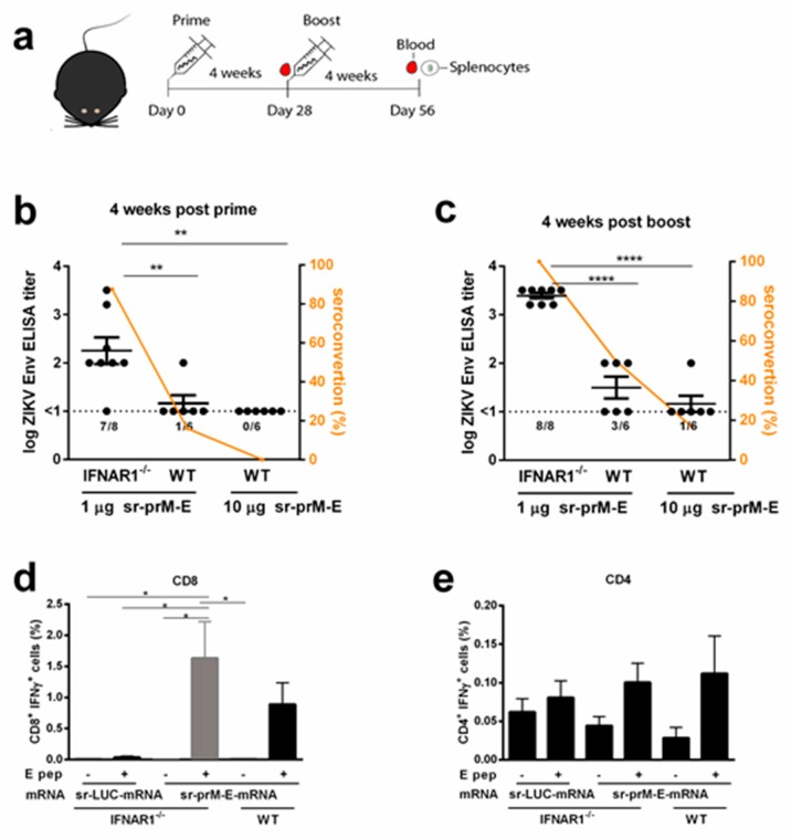
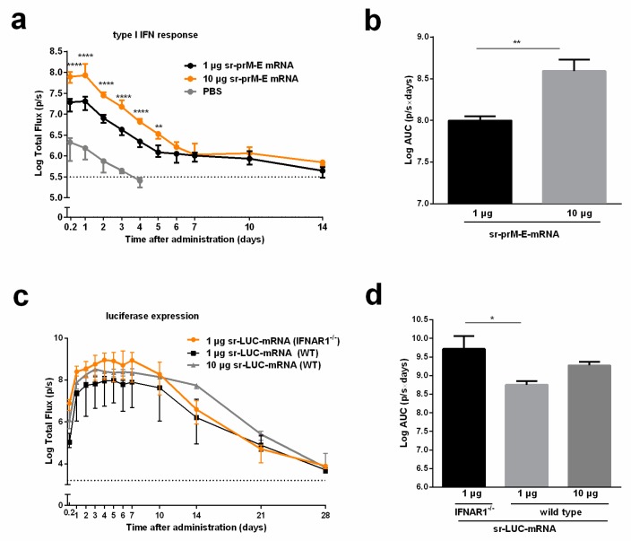
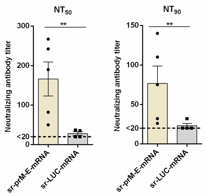
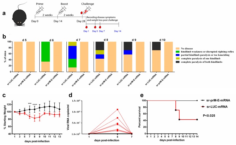
To combat emerging infectious diseases like Zika virus (ZIKV), synthetic messenger RNAs (mRNAs) encoding viral antigens are very attractive as they allow a rapid, generic, and flexible production of vaccines. In this work, we engineered a self-replicating mRNA (sr-mRNA) vaccine encoding the pre-membrane and envelope (prM-E) glycoproteins of ZIKV. Intradermal electroporation of as few as 1 µg of this mRNA-based ZIKV vaccine induced potent humoral and cellular immune responses in BALB/c and especially IFNAR1-/- C57BL/6 mice, resulting in a complete protection of the latter mice against ZIKV infection. In wild-type C57BL/6 mice, the vaccine resulted in very low seroconversion rates and antibody titers. The potency of the vaccine was inversely related to the dose of mRNA used in wild-type BALB/c or C57BL/6 mice, as robust type I interferon (IFN) response was determined in a reporter mice model (IFN-β+/Δβ-luc). We further investigated the inability of the sr-prM-E-mRNA ZIKV vaccine to raise antibodies in wild-type C57BL/6 mice and found indications that type I IFNs elicited by this naked sr-mRNA vaccine might directly impede the induction of a robust humoral response. Therefore, we assume that the efficacy of sr-mRNA vaccines after intradermal electroporation might be increased by strategies that temper their inherent innate immunogenicity.

Keywords: IFNAR1 knockout mice; Zika virus vaccine; self-replicating mRNA; type I interferon response.


Conflict of interest statement

The authors declare no conflict of interest.

Figures

Figure 1
Figure 1
Construction and characterization of a self-replicating (sr) mRNA vaccine against Zika virus (ZIKV). (a) Depiction of the sr pre-membrane and envelope (prM-E) mRNA ZIKV vaccine with the Japanese encephalitis virus (JEV) signal sequence in front of the prM-E, and the control sr-mRNA encoding luciferase (sr-LUC-mRNA). The JEV-prM-E and luciferase sequences were codon-optimized. The replication of the sr-mRNA is mediated by the four nonstructural proteins (nsPs) of Venezuelan equine encephalitis virus (VEEV). The UTRs and the subgenomic promotor (SGP) are also derived from VEEV. After transfection of the sr-prM-E-mRNA in baby hamster kidney (BHK)-21 cells, the expression of the ZIKV E protein (~54 kDa) in the cells was detected by Western blot (b) and its secretion in supernatant by dot blot (c) using monoclonal antibodies against ZIKV E protein and denaturing conditions. BHK cells transfected with sr-LUC-mRNA served as negative controls.
Figure 2
Figure 2
Immunogenicity of the sr-prM-E-mRNA and FI-ZIKV vaccines in BALB/c mice. (a) Mice were intradermally electroporated on day 0 and 28 with either 1 or 10 μg of sr-prM-E-mRNA ZIKV vaccine, 1 µg of FI-ZIKV vaccine, or with 1 μg sr-LUC-mRNA. Antibody titers were determined four weeks after the prime (b) or boost (c) by a ZIKV E protein-specific IgG ELISA (n = 12 in each group). The dashed lines indicate the limit of detection of the assay. The lower panels depict ZIKV E protein-specific CD8+ (d) and CD4+ (e) T cell responses in mice vaccinated with 1 µg of sr-prM-E mRNA vaccine (n = 5 in each group). Mice vaccinated with either 1 µg sr-LUC-mRNA served as negative controls (n = 5). T cell responses were determined four weeks after the boost by intracellular staining of IFN-γ in T cells stimulated with a ZIKV E protein peptide pool (E pep +). Mice receiving only alum adjuvant served as controls for the FI-ZIKV vaccinated mice. These mice did not develop ZIKV E protein-specific antibodies or cellular immune responses, as observed for the sr-LUC-mRNA control mice (data not shown). Data are represented as mean ± SEM and were analyzed by ANOVA followed by Tukey’s test.
Figure 3
Figure 3
Immunogenicity of the sr-prM-E mRNA vaccine in IFNAR1-/- mice. (a) C57BL/6 wild-type (WT) and IFNAR1-/- mice were intradermally electroporated on day 0 and 28 with 1 μg of sr-prM-E-mRNA ZIKV vaccine. An additional group of WT mice was vaccinated in a similar way with a tenfold higher dose of the vaccine. Antibody titers in IFNAR1-/- and WT mice were determined four weeks after the prime (b) or boost (c) by a ZIKV E-protein specific IgG ELISA (n = 6 or 8). The dashed lines indicate the limit of detection of the assay. The lower panels show ZIKV E-protein specific CD8+ (d) and CD4+ (e) T cell responses in IFNAR1-/- and WT mice receiving 1 µg sr-prM-E mRNA vaccine or sr-LUC-mRNA. T cell responses were determined four weeks after the boost by intracellular staining of IFN-γ in T cells stimulated with a ZIKV E protein peptide pool (E pep +) (n = 6 or 8). All data are represented as mean ± SEM and were analyzed by ANOVA followed by Tukey’s test.
Figure 4
Figure 4
Interferon response after intradermal electroporation of the sr-prM-E mRNA ZIKV vaccine and effect of IFNAR1 on the expression of sr-LUC-mRNA. (a) IFN-β luciferase reporter (IFN-β+/Δβ-luc) mice were intradermally electroporated with 1 or 10 μg of sr-prM-E-mRNA ZIKV vaccine and the type I interferon response was monitored by measuring the bioluminescent signal at the injection spot for 14 days. The AUC of the curves in (a) are shown in (b) (n = 3). (c) C57BL/6 WT and IFNAR1-/- mice were intradermally electroporated with 1 or 10 μg sr-LUC-mRNA and the luciferase expression was determined by measuring the bioluminescent signal at the injection spot for 28 days. The results of the statistical analysis of the data shown in (c) can be found Table S2. The AUC of the curves in (c) are shown in (d) (n = 4). Data are represented as median with interquartile range and statistical analysis was performed using ANOVA followed by Tukey’s test.
Figure 5
Figure 5
Zika virus neutralizing antibody titers after vaccination of IFNAR1-/- mice with the sr-prM-E mRNA vaccine. Mice were intradermally electroporated on day 0 and 14 with 1 μg of sr-prM-E-mRNA ZIKV vaccine or sr-LUC-mRNA and two weeks after the boost, ZIKV-specific neutralizing antibody titers were determined by a neutralization test (NT). The neutralizing antibody titers are expressed as the reciprocal of the endpoint serum dilution that neutralized the challenge virus by (a) 50% (NT50) or (b) 90% (NT90). The lower limit of NT quantification was 20 and is represented by the dashed line (n = 5 sr-prM-E-mRNA group and n = 4 sr-LUC-mRNA group).
Figure 6
Figure 6
Protective efficacy of the sr-prM-E mRNA vaccine in IFNAR1-/- mice. (a) Mice were intradermally electroporated on day 0 and 14 with 1 μg of sr-prM-E-mRNA ZIKV vaccine or sr-LUC-mRNA. Two weeks after the boost they were challenged by injecting 1000 TCID50 of ZIKV (MR-766) in the footpad (n = 7); (b) The disease symptoms were daily recorded from day 5 to day 10 after ZIKV challenge and (c) the percentage of weight loss relative to the body mass just before ZIKV infection was calculated; (d) the ZIKV RNA loads in plasma were determined by qRT-PCR. The dashed line indicates the limit of detection of the assay; (e) survival rates were determined based on the scoring system described in Table S1.

References

    1. Johansson M.A., Mier-y-Teran-Romero L., Reefhuis J., Gilboa S.M., Hills S.L. Zika and the Risk of Microcephaly. N. Engl. J. Med. 2016;375:1–4. doi: 10.1056/NEJMp1605367. - DOI - PMC - PubMed
    1. Nambala P., Su W.C. Role of Zika Virus prM Protein in Viral Pathogenicity and Use in Vaccine Development. Front. Microbiol. 2018;9:1797. doi: 10.3389/fmicb.2018.01797. - DOI - PMC - PubMed
    1. Besnard M., Lastere S., Teissier A., Cao-Lormeau V., Musso D. Evidence of perinatal transmission of Zika virus, French Polynesia, December 2013 and February 2014. Eurosurveillance. 2014;19:20751. doi: 10.2807/1560-7917.ES2014.19.13.20751. - DOI - PubMed
    1. Oehler E., Watrin L., Larre P., Leparc-Goffart I., Lastere S., Valour F., Baudouin L., Mallet H., Musso D., Ghawche F. Zika virus infection complicated by Guillain-Barre syndrome—Case report, French Polynesia, December 2013. Eurosurveillance. 2014;19:20720. doi: 10.2807/1560-7917.ES2014.19.9.20720. - DOI - PubMed
    1. Oehler E., Fournier E., Leparc-Goffart I., Larre P., Cubizolle S., Sookhareea C., Lastere S., Ghawche F. Increase in cases of Guillain-Barre syndrome during a Chikungunya outbreak, French Polynesia, 2014 to 2015. Eurosurveillance. 2015;20:30079. doi: 10.2807/1560-7917.ES.2015.20.48.30079. - DOI - PubMed