Review
. 2019 Aug 23;8(9):963.
doi: 10.3390/cells8090963.

Signaling Pathways of Type I and Type III Interferons and Targeted Therapies in Systemic Lupus Erythematosus

Affiliations
Review

Signaling Pathways of Type I and Type III Interferons and Targeted Therapies in Systemic Lupus Erythematosus

I-Tsu Chyuan et al. Cells. .

Abstract

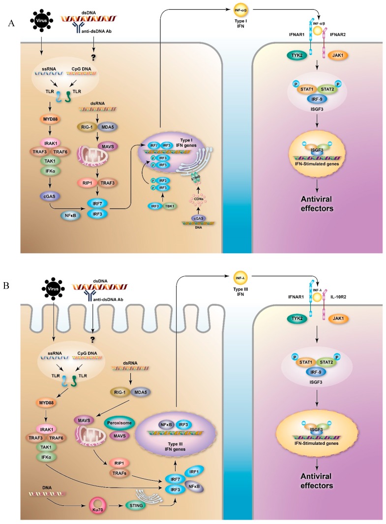
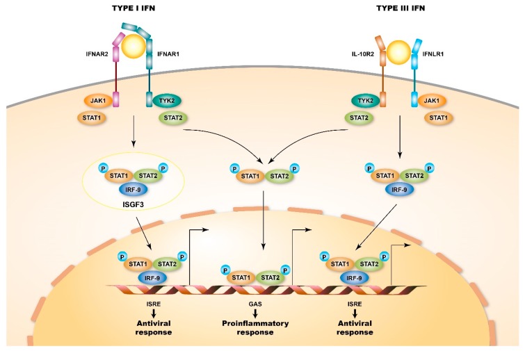
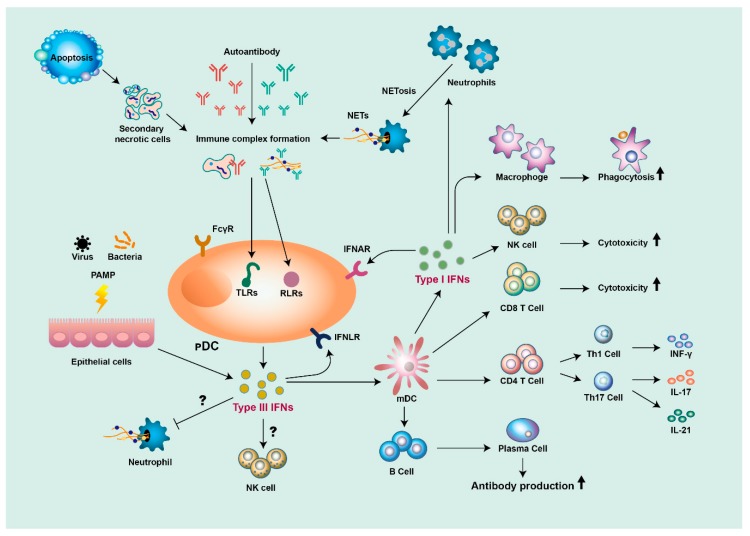
Type I and type III interferons (IFNs) share several properties in common, including the induction of signaling pathways, the activation of gene transcripts, and immune responses, against viral infection. Recent advances in the understanding of the molecular basis of innate and adaptive immunity have led to the re-examination of the role of these IFNs in autoimmune diseases. To date, a variety of IFN-regulated genes, termed IFN signature genes, have been identified. The expressions of these genes significantly increase in systemic lupus erythematosus (SLE), highlighting the role of type I and type III IFNs in the pathogenesis of SLE. In this review, we first discussed the signaling pathways and the immunoregulatory roles of type I and type III IFNs. Next, we discussed the roles of these IFNs in the pathogenesis of autoimmune diseases, including SLE. In SLE, IFN-stimulated genes induced by IFN signaling contribute to a positive feedback loop of autoimmunity, resulting in perpetual autoimmune inflammation. Based on this, we discussed the use of several specific IFN blocking strategies using anti-IFN-α antibodies, anti-IFN-α receptor antibodies, and IFN-α-kinoid or downstream small molecules, which intervene in Janus kinase (JAK)-signal transducer and activator of transcription (STAT) pathways, in clinical trials for SLE patients. Hopefully, the development of novel regimens targeting IFN signaling pathways will shed light on promising future therapeutic applications for SLE patients.

Keywords: T cell; dendritic cell; interferon; interferon receptor signaling; systemic lupus erythematosus.


Conflict of interest statement

The authors have no conflicts of interest to declare.

Figures

Figure 1
Figure 1
Signaling pathways of type I and type III interferons (IFNs). The production of (A) type I and (B) type III IFNs can be induced by virus infection or by immune complexes, which are sensed by pattern recognition receptors (PRRs), especially toll-like receptors (TLRs) and retinoic acid-inducible gene 1 (RIG-I)-like receptors (RLRs). Differential signaling molecules lead to the activation of the transcription factors nuclear factor kappa-light-chain-enhancer of activated B cells (NF-κB) and interferon regulatory factors (IRFs), and eventually to the activation of IFN gene transcripts. The secreted IFNs ligate to the type I or type III IFN receptors (IFN-α/β receptor (IFNAR)1/IFNAR2 or IFN-λ receptor (IFNLR)1/interleukin-10 receptor (IL-10R)2, respectively) of the neighboring cells and stimulate the production of IFN-stimulated genes (ISGs) via Janus kinase (JAK)-signal transducer and activator of transcription (STAT) pathways, which results in the production of several antiviral effectors. Cyclic GMP-AMP synthase (cGAS) transforms DNA into cyclic di-nucleotides (CDNs), which can be recognized by stimulator of interferon genes (STING). STING triggers the activation of tank-binding kinase 1 (TBK1) to phosphorylate IRF-3 and induces the transcription of type I IFNs. In the transcription of type III IFNs, the activation of STING is involved in Ku70.
Figure 2
Figure 2
Signaling pathways of type I and type III IFN receptors. Type I and type III IFN receptors are heterodimers and consist of different receptor chains (IFNAR1 and IFNAR2 for the type I IFN receptor and IFNLR1 and IL10R2 for the type III IFN receptor). Both receptors are associated with two kinases from the JAK family: JAK1 and TYK2 (tyrosine kinase 2). When ligating to their cognate ligand, JAK kinases auto-phosphorylate the IFN receptor, which results in the recruitment of signal transducer and activator of transcription (STAT) proteins, phosphorylation, dimerization, and nuclear translocation. In particular, STAT1, STAT2, and IFN-regulatory factor 9 (IRF9) form interferon-stimulated gene factor 3 (ISGF3) complexes and bind to IFN-stimulated response element (ISRE) sequences, thus activating classical antiviral genes. Additionally, STAT1 homodimers bind to gamma-activated sequences (GASs), thus inducing the activation of pro-inflammatory genes.
Figure 3
Figure 3
The roles of type I and type III IFNs in the pathogenesis of SLE (systemic lupus erythematosus). Self-nucleic acid from apoptotic cells, or from neutrophil extracellular traps (NETs) released from neutrophils, are detected by autoantibodies to form immune complexes, stimulating plasmacytoid dendritic cells (pDCs) to produce type I and type III IFNs. Epithelial cells also produce type III IFNs in response to pathogen-associated molecular patterns (PAMPs). Both type I and type III IFNs stimulate myeloid DCs (mDCs) to activate T- and B-cells, which leads to the production of diverse proinflammatory cytokines and autoantibodies. Type I IFNs can also promote cytotoxicity function in macrophages and natural killer (NK) cells. Both type I and type III IFNs can contribute to a positive feedback loop of inflammation.

References

    1. Feldman C.H., Hiraki L.T., Liu J., Fischer M.A., Solomon D.H., Alarcon G.S., Winkelmayer W.C., Costenbader K.H. Epidemiology and sociodemographics of systemic lupus erythematosus and lupus nephritis among US adults with Medicaid coverage, 2000–2004. Arthritis Rheum. 2013;65:753–763. doi: 10.1002/art.37795. - DOI - PMC - PubMed
    1. Taylor H.G., Stein C.M. Systemic lupus erythematosus in Zimbabwe. Ann. Rheum. Dis. 1986;45:645–648. doi: 10.1136/ard.45.8.645. - DOI - PMC - PubMed
    1. Rees F., Doherty M., Grainge M.J., Lanyon P., Zhang W. The worldwide incidence and prevalence of systemic lupus erythematosus: A systematic review of epidemiological studies. Rheumatology (Oxford) 2017;56:1945–1961. doi: 10.1093/rheumatology/kex260. - DOI - PubMed
    1. Gorji A.E., Roudbari Z., Alizadeh A., Sadeghi B. Investigation of systemic lupus erythematosus (SLE) with integrating transcriptomics and genome wide association information. Gene. 2019;706:181–187. doi: 10.1016/j.gene.2019.05.004. - DOI - PubMed
    1. Deng Y., Tsao B.P. Updates in Lupus Genetics. Curr. Rheumatol. Rep. 2017;19:68. doi: 10.1007/s11926-017-0695-z. - DOI - PubMed

Publication types

MeSH terms