. 2019 Oct 11;125(9):805-820.
doi: 10.1161/CIRCRESAHA.119.315082. Epub 2019 Aug 27.

HIV-Nef Protein Transfer to Endothelial Cells Requires Rac1 Activation and Leads to Endothelial Dysfunction Implications for Statin Treatment in HIV Patients

Affiliations

HIV-Nef Protein Transfer to Endothelial Cells Requires Rac1 Activation and Leads to Endothelial Dysfunction Implications for Statin Treatment in HIV Patients

Sarvesh Chelvanambi et al. Circ Res. .

Abstract

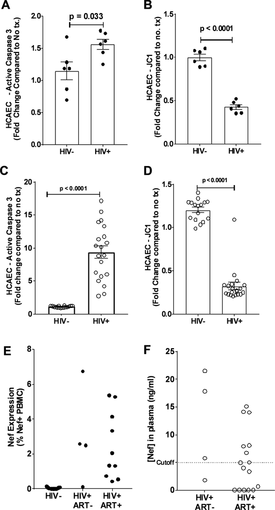
Rationale: Even in antiretroviral therapy-treated patients, HIV continues to play a pathogenic role in cardiovascular diseases. A possible cofactor may be persistence of the early HIV response gene Nef, which we have demonstrated recently to persist in the lungs of HIV+ patients on antiretroviral therapy. Previously, we have reported that HIV strains with Nef, but not Nef-deleted HIV strains, cause endothelial proinflammatory activation and apoptosis.

Objective: To characterize mechanisms through which HIV-Nef leads to the development of cardiovascular diseases using ex vivo tissue culture approaches as well as interventional experiments in transgenic murine models.

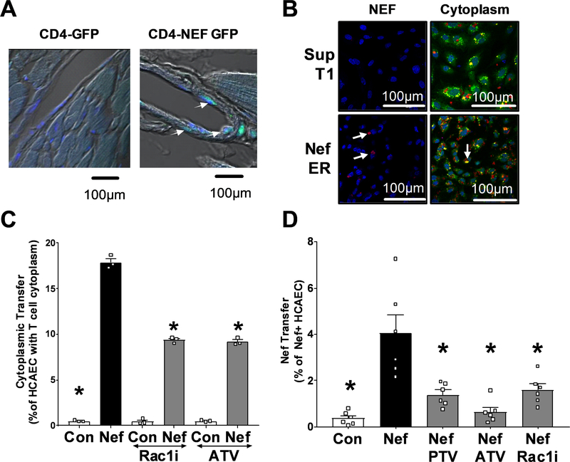
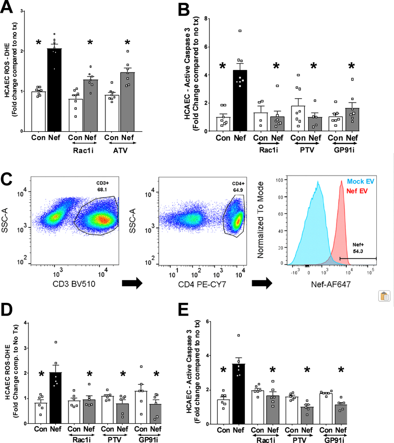
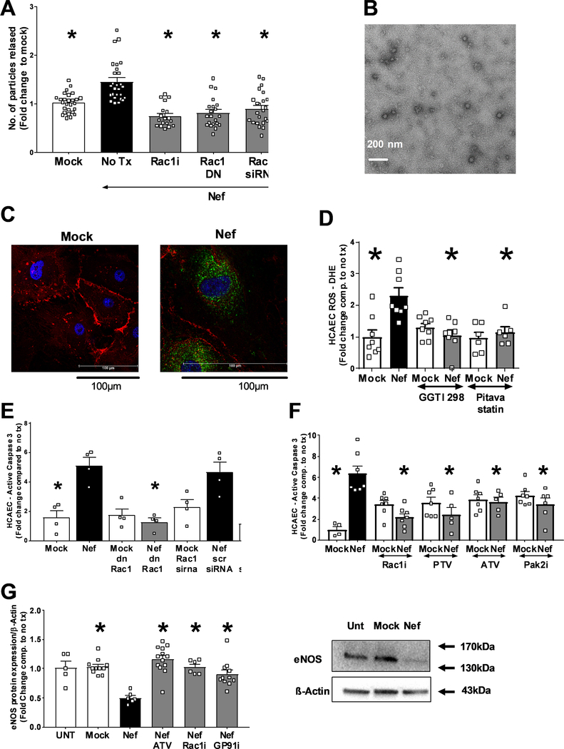
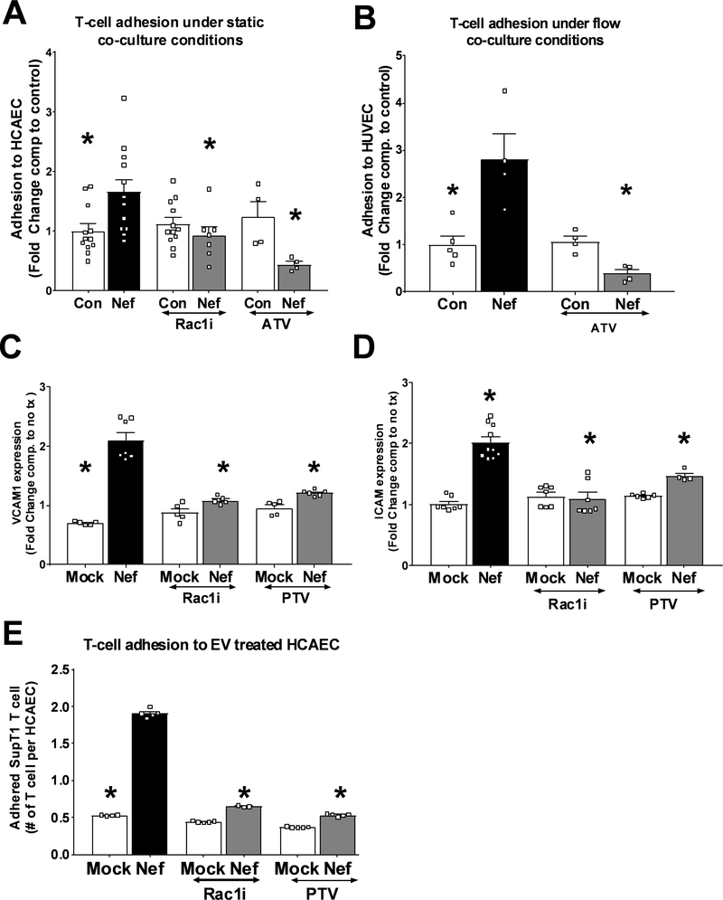
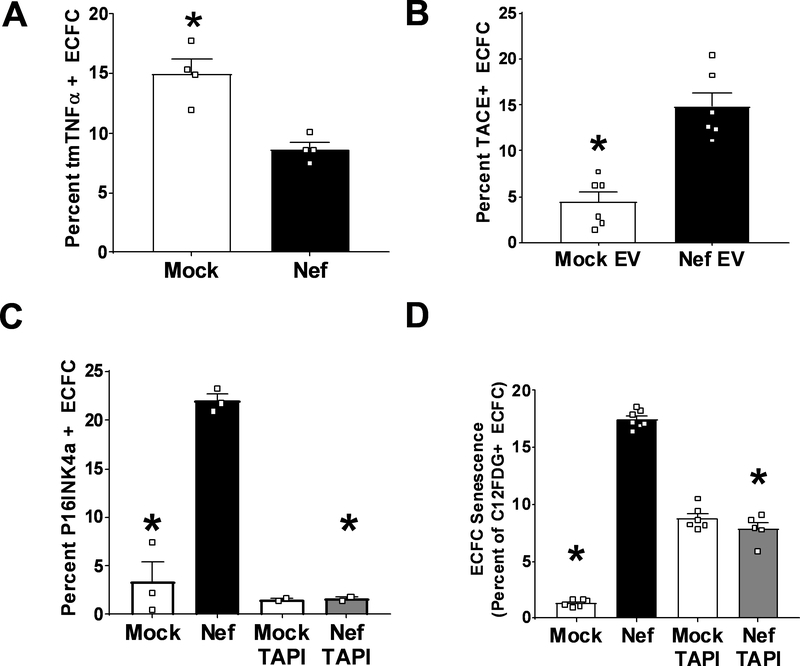
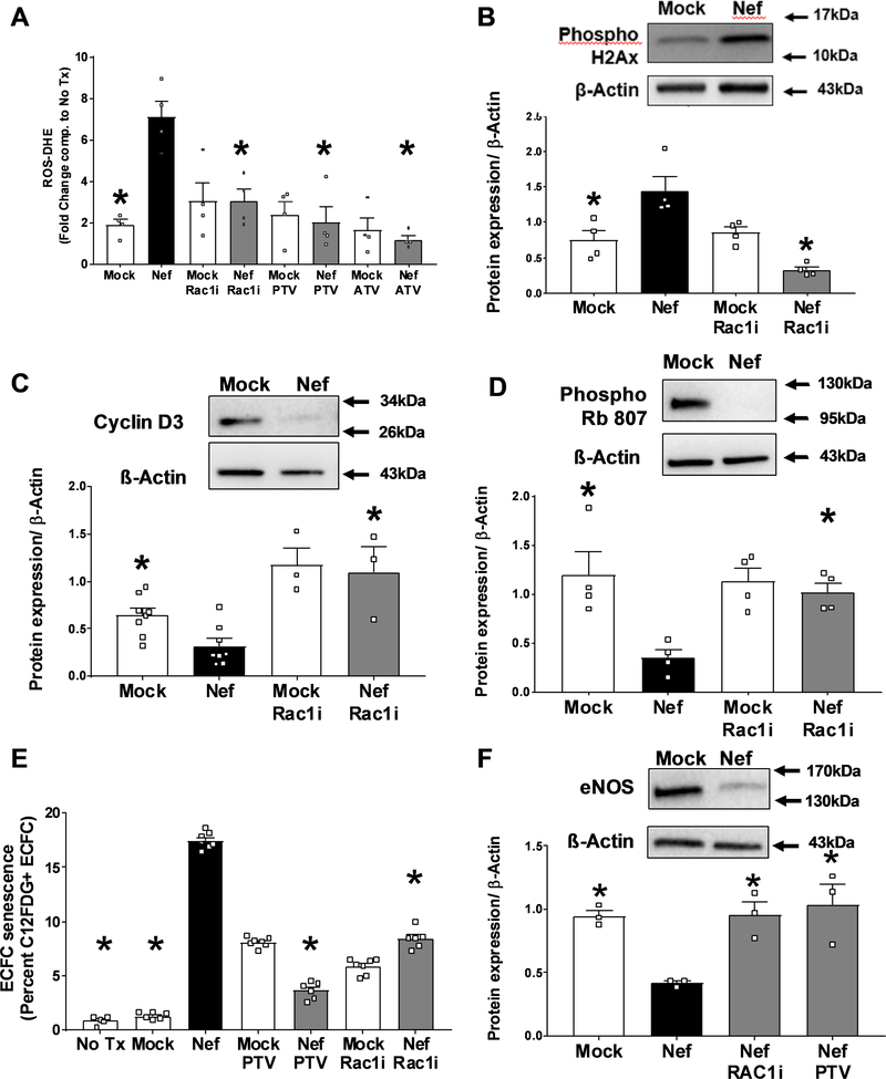
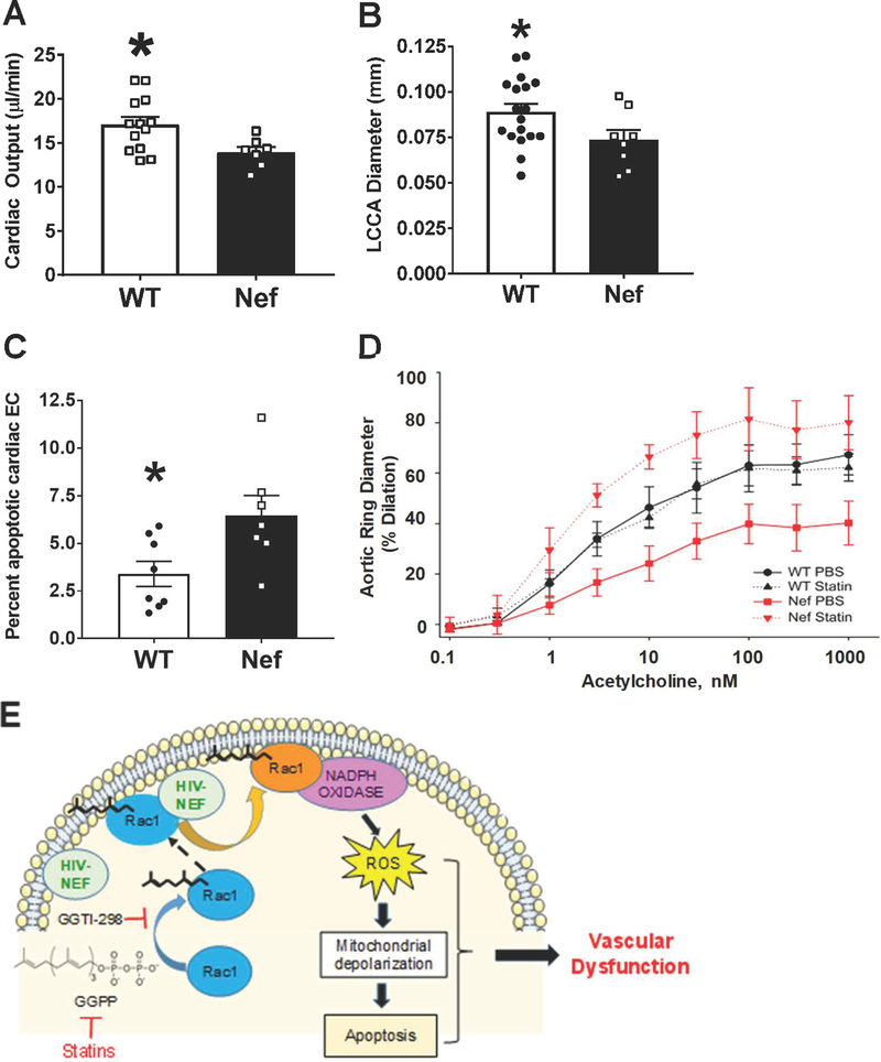
Methods and results: Extracellular vesicles derived from both peripheral blood mononuclear cells and plasma from HIV+ patient blood samples induced human coronary artery endothelial cells dysfunction. Plasma-derived extracellular vesicles from antiretroviral therapy+ patients who were HIV-Nef+ induced significantly greater endothelial apoptosis compared with HIV-Nef-plasma extracellular vesicles. Both HIV-Nef expressing T cells and HIV-Nef-induced extracellular vesicles increased transfer of cytosol and Nef protein to endothelial monolayers in a Rac1-dependent manner, consequently leading to endothelial adhesion protein upregulation and apoptosis. HIV-Nef induced Rac1 activation also led to dsDNA breaks in endothelial colony forming cells, thereby resulting in endothelial colony forming cell premature senescence and endothelial nitric oxide synthase downregulation. These Rac1-dependent activities were characterized by NOX2-mediated reactive oxygen species production. Statin treatment equally inhibited Rac1 inhibition in preventing or reversing all HIV-Nef-induction abnormalities assessed. This was likely because of the ability of statins to block Rac1 prenylation as geranylgeranyl transferase inhibitors were effective in inhibiting HIV-Nef-induced reactive oxygen species formation. Finally, transgenic expression of HIV-Nef in endothelial cells in a murine model impaired endothelium-mediated aortic ring dilation, which was then reversed by 3-week treatment with 5 mg/kg atorvastatin.

Conclusions: These studies establish a mechanism by which HIV-Nef persistence despite antiretroviral therapy could contribute to ongoing HIV-related vascular dysfunction, which may then be ameliorated by statin treatment.

Keywords: apoptosis; cardiovascular diseases; endothelial progenitor cells; extracellular vesicles; statin.


Figures

Figure 1:
Figure 1:. HIV-Nef protein persists in virologically suppressed HIV+ patients.
Addition of PBMCs (A and B, closed circles) and extracellular vesicles (C and D, open circles) from HIV+ patients to HCAEC induced endothelial cell apoptosis as measured by active caspase 3 levels (A and B) and mitochondrial depolarization using JC-1 (C and D). HIV-Nef protein persists in PBMCs (E) and plasma (F) of HIV+ patients either treatment naïve or on antiretroviral therapy. Dotted line indicates cutoff (5ng/ml) based on HIV- control plasma and previously published results. Statistical significance between groups was determined using Student’s T-Test with Welch’s correction. Raw p-value is indicated in graphs.
Figure 2:
Figure 2:. HIV-Nef protein promotes cytoplasmic transfer from T cells to coronary artery endothelial cells.
Heart sections of single CD4-GFP and double CD4-Nef-GFP transgenic mice were stained with αGFP mab (green). White arrows indicate T-Cell cytoplasm within endothelial lining (A). HIV-Nef protein transfer from SupT1 or Nef-ER T cells to HCAEC is visualized using anti-Nef EH1 mAB (left panels). Cytoplasmic transfer of Cell Tracker Deep Red Dye from T cells is seen in Calcein AM stained endothelial cells (right panels) (B). Quantification of cytoplasmic transfer (C) or HIV-Nef protein transfer (D) from T-Cells to HCAEC was quantified using FACS. ATV=5μmol/l atorvastatin; PTV=100nmol/l Pitavastatin; Rac1i = 5μmol/l NSC23766. Groups were compared using one-way ANOVA followed by Tukey Post-hoc test. * denotes adjusted p-value<0.001 compared to HIV-Nef group (black bar). Scale bars denote 100um.
Figure 3:
Figure 3:. Rac1 inhibition and statin treatment block HIV-Nef protein induced endothelial cell stress.
Co-culture of HIV-Nef expressing T cells (Nef) induced ROS production (A) and apoptosis (B) in HCAEC. HIV-Nef protein was taken up by CD45+/CD3+/CD4+ T cells when PBMC were treated with mock ev or HIV-Nef EV (Nef) for 2hr (Blue= mock, Red = Nef) (C). HIV-Nef EV treated PBMC induced ROS production (D) and caspase 3 activation (E) in HCAEC. ATV=5μmol/l atorvastatin; PTV=100nmol/l Pitavastatin; Rac1i = 5μmol/l NSC23766. GP91i = Inhibitory peptide 1μmol/l. Groups were compared using one-way ANOVA followed by Tukey Post-hoc test. * denotes adjusted p-value<0.001 compared to HIV-Nef group (black bar).
Figure 4:
Figure 4:. HIV-Nef EV induce endothelial cell apoptosis.
(A) HIV-Nef transfection into HEK293T increases the amount of EV released. EV morphology was analyzed using transmission electron microscopy (B) with scale bar denoting 200nm. (C) Addition of HIV-Nef+ EV transferred HIV-Nef protein to HCAEC (Green = αNef EH1mab; Red = PECAM; Blue = DAPI; Scale bars = 100 μm). HIV-Nef+ EV promotes ROS production (D), apoptosis (E and F) and downregulation of eNOS (G) in HCAEC. ATV=5μmol/l atorvastatin; PTV=100nmol/l pitavastatin; Rac1i = 5μmol/l NSC23766. GP91i = Inhibitory peptide 1μmol/l; Pak2i = 5μmol/l FRAX597 GGTI-298 = 5μmol/l. Groups were compared using one-way ANOVA followed by Tukey Post-hoc test. * denotes adjusted p-value<0.001 compared to HIV-Nef group (black bar) # denotes p-value<0.0001 compared to HIV-Nef scr siRNA.
Figure 5:
Figure 5:. HIV-Nef expression enhances interaction between T-cell and endothelial cells.
Compared to control SupT1 T cells (SupT1), HIV-Nef expressing T cells (Nef-ER) displayed increased adhesion to HCAEC under static (A) and to HUVEC under flow (B) conditions. HCAEC treated 24hr with 3μg/ml Nef EV showed increased VCAM1 (C) and ICAM1 (D) surface expression as determined by FACS. T-cell adhesion to HCAEC pre-treated with 3μg/ml of mock ev or HIV-Nef+ EV in the presence of Rac1 inhibitors (E). ATV=5μmol/l atorvastatin; PTV=100nmol/l pitavastatin; Rac1i = 5μmol/l NSC23766. Groups were compared using one-way ANOVA followed by Tukey Post-hoc test. * denotes adjusted p-value <0.001 compared to HIV-Nef group (black bar).
Figure 6:
Figure 6:. HIV-Nef EV downregulates tmTNFα to induce premature ECFC senescence.
Addition of HIV-Nef+ EV downregulates surface expression of transmembrane TNFα (A) via upregulation of TACE activity (B). HIV-Nef+ EV induced ECFC senescence as evidenced by p16INK4a induction (C) and lysosomal β-galactosidase activity (D). TAPI = 10μmol/l. Groups were compared using one-way ANOVA followed by Tukey Post-hoc test or Student’s t-test with Welch’s correction. * denotes adjusted p-value <0.05 compared to HIV-Nef group (black bar).
Figure 7:
Figure 7:. HIV-Nef+ EV induce ECFC dysfunction.
Addition of HIV-Nef EV induce ROS production (A) and double stranded DNA breaks (B) in ECFC quantified using DHE staining and phosphorylated histone 2Ax respectively. HIV-Nef+ EV downregulates of Cyclin D3 (C) and Phospho Rb 807 (D). HIV-Nef+ EV induces premature senescence in ECFC quantified using C12FDG staining to measure lysosomal β-galactosidase activity (E). HIV-Nef+ EV downregulates eNOS protein expression in ECFC (F). ATV=5μmol/l atorvastatin; PTV=100nmol/l pitavastatin; Rac1i = 5μmol/l NSC23766. Groups were compared using one-way ANOVA followed by Tukey Post-hoc test or Student’s t-test with Welch’s correction. * denotes adjusted p-value <0.05 compared to HIV-Nef group (black bar).
Figure 8:
Figure 8:. Endothelial expression of HIV-Nef in VE-Cadherin promoter driven HIV-Nef transgenic mice induces cardiovascular dysfunction.
Echocardiography was used to evaluate Left Circumflex Coronary Artery (LCCA) diameter (A) and cardiac output (B). Endothelial cells in the heart showed increased apoptosis measured through cleaved caspase 3 staining in CD31+/CD45- endothelial cells (C). Aortic rings isolated from WT and HIV-Nef expressing mice treated with vehicle or 5mg/kg atorvastatin (daily for 3 weeks) were preconstricted using phenylephrine and endothelium dependent vasodilation was measured in response to increasing concentration of acetylcholine (D). Schematic describes proposed mechanism of HIV-Nef induced endothelial dysfunction (E). Groups were compared using Student’s t-test with Welch’s correction. * denotes adjusted p-value<0.05 compared to HIV-Nef group.

Comment in

References

    1. Paisible AL, Chang CC, So-Armah KA, Butt AA, Leaf DA, Budoff M, Rimland D, Bedimo R, Goetz MB, Rodriguez-Barradas MC, Crane HM, Gibert CL, Brown ST, Tindle HA, Warner AL, Alcorn C, Skanderson M, Justice AC, Freiberg MS. Hiv infection, cardiovascular disease risk factor profile, and risk for acute myocardial infarction. J Acquir Immune Defic Syndr. 2015;68:209–216 - PMC - PubMed
    1. Beckman JA, Duncan MS, Alcorn CW, So-Armah K, Butt AA, Goetz MB, Tindle HA, Sico J, Tracy RP, Justice AC, Freiberg MS. Association of hiv infection and risk of peripheral artery disease. Circulation. 2018 - PMC - PubMed
    1. Francisci D, Giannini S, Baldelli F, Leone M, Belfiori B, Guglielmini G, Malincarne L, Gresele P. Hiv type 1 infection, and not short-term haart, induces endothelial dysfunction. AIDS. 2009;23:589–596 - PubMed
    1. Currier JS, Lundgren JD, Carr A, Klein D, Sabin CA, Sax PE, Schouten JT, Smieja M. Epidemiological evidence for cardiovascular disease in hiv-infected patients and relationship to highly active antiretroviral therapy. Circulation. 2008;118:e29–35 - PMC - PubMed
    1. Triant VA, Lee H, Hadigan C, Grinspoon SK. Increased acute myocardial infarction rates and cardiovascular risk factors among patients with human immunodeficiency virus disease. J Clin Endocrinol Metab. 2007;92:2506–2512 - PMC - PubMed

Publication types

MeSH terms

Substances