Review
. 2020 Feb 3;12(2):a037192.
doi: 10.1101/cshperspect.a037192.

Epicardium in Heart Development

Affiliations
Review

Epicardium in Heart Development

Yingxi Cao et al. Cold Spring Harb Perspect Biol. .

Abstract

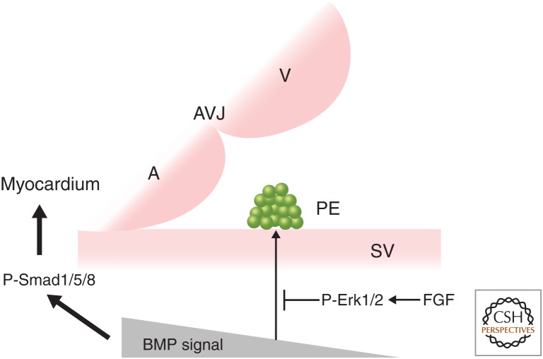
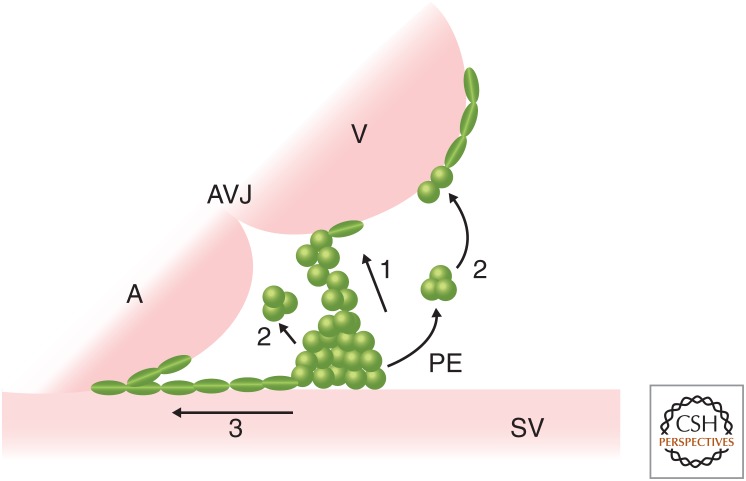
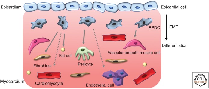
The epicardium, the outermost tissue layer that envelops all vertebrate hearts, plays a crucial role in cardiac development and regeneration and has been implicated in potential strategies for cardiac repair. The heterogenous cell population that composes the epicardium originates primarily from a transient embryonic cell cluster known as the proepicardial organ (PE). Characterized by its high cellular plasticity, the epicardium contributes to both heart development and regeneration in two critical ways: as a source of progenitor cells and as a critical signaling hub. Despite this knowledge, there are many unanswered questions in the field of epicardial biology, the resolution of which will advance the understanding of cardiac development and repair. We review current knowledge in cross-species epicardial involvement, specifically in relation to lineage specification and differentiation during cardiac development.


Figures

Figure 1.
Figure 1.
Bone morphogenetic protein (BMP) and fibroblast growth factor (FGF) signals are involved in the induction of the proepicardial organ (PE). In chick embryos, strong BMP signaling promotes myocardial differentiation of the precursor pool of the splanchnic mesoderm through p-Smad1/5/8, whereas weak BMP signaling stimulates PE formation. FGF signaling negatively regulates BMP signaling in the PE region via p-Erk1/2 to promote PE formation, although whether FGF signaling is required for the induction or maintained expression of PE-specific genes, such as Tbx18, is controversial (van Wijk et al. 2009; Torlopp et al. 2010). In contrast, blocking BMP signaling in zebrafish embryos reduces tbx18 and tcf21 expression and PE specification, whereas elevated bmp2b expression before PE formation induces ectopic expression of tbx18 (Liu and Stainier 2010). A, Atrium; V, ventricle; AVJ, atrioventricular junction; SV, sinus venosus.
Figure 2.
Figure 2.
An overview of proepicardial organ (PE) translocation to the myocardium. Schematic of PE translocation to the myocardium showing three models across species. (1) Direct contact: In the chick, Xenopus, axolotl, zebrafish, and mouse, the PE produces protrusions to form a tissue bridge between the PE and the dorsal wall of the ventricle (or the PE contacts the ventricle directly) to allow the PE cells to migrate on to the myocardium. (2) Floating cysts: In zebrafish and mice, PE cell clusters detach from the proepicardial surface to form cysts, which float across the pericardial cavity to attach to the ventricular wall. (3) Migration: In mice, PE cells migrate from the SV toward the heart along the surface of the inflow tract. A, Atrium; V, ventricle; AVJ, atrioventricular junction; SV, sinus venosus.
Figure 3.
Figure 3.
Cellular contributions of epicardium during heart development. Epicardial cells enter the myocardium through epithelial-to-mesenchymal transition (EMT) to form epicardium-derived cells (EPDCs), which differentiate into fibroblasts, vascular smooth muscle cells (vSMCs), pericytes, fat cells, and, possibly, endothelial cells and cardiomyocytes. Solid arrows denote consensus cell fates, whereas dashed arrows indicate controversial contributions.

Similar articles

Cited by

References

    1. Acharya A, Baek ST, Huang G, Eskiocak B, Goetsch S, Sung CY, Banfi S, Sauer MF, Olsen GS, Duffield JS, et al. 2012. The bHLH transcription factor Tcf21 is required for lineage-specific EMT of cardiac fibroblast progenitors. Development 139: 2139–2149. 10.1242/dev.079970 - DOI - PMC - PubMed
    1. Ali SR, Ranjbarvaziri S, Talkhabi M, Zhao P, Subat A, Hojjat A, Kamran P, Muller AM, Volz KS, Tang Z, et al. 2014. Developmental heterogeneity of cardiac fibroblasts does not predict pathological proliferation and activation. Circ Res 115: 625–635. 10.1161/CIRCRESAHA.115.303794 - DOI - PubMed
    1. Andrés-Delgado L, Ernst A, Galardi-Castilla M, Bazaga D, Peralta M, Münch J, González-Rosa JM, Marques I, Tessadori F, de la Pompa JL, et al. 2019. Actin dynamics and the Bmp pathway drive apical extrusion of proepicardial cells. Development 146: dev174961 10.1242/dev.174961 - DOI - PMC - PubMed
    1. Antonopoulos AS, Antoniades C. 2017. The role of epicardial adipose tissue in cardiac biology: classic concepts and emerging roles. J Physiol 595: 3907–3917. 10.1113/JP273049 - DOI - PMC - PubMed
    1. Austin AF, Compton LA, Love JD, Brown CB, Barnett JV. 2008. Primary and immortalized mouse epicardial cells undergo differentiation in response to TGFβ. Dev Dyn 237: 366–376. 10.1002/dvdy.21421 - DOI - PubMed

Publication types

LinkOut - more resources