Review
. 2020 Feb 3;12(2):a036434.
doi: 10.1101/cshperspect.a036434.

Cell Death and Neurodegeneration

Affiliations
Review

Cell Death and Neurodegeneration

Benjamin J Andreone et al. Cold Spring Harb Perspect Biol. .

Abstract

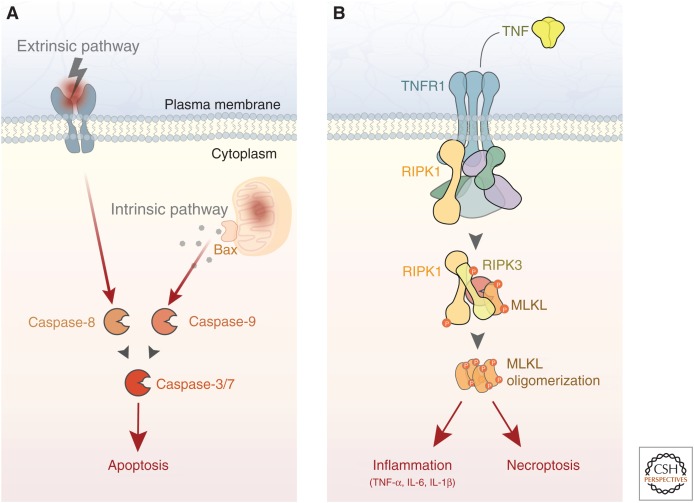
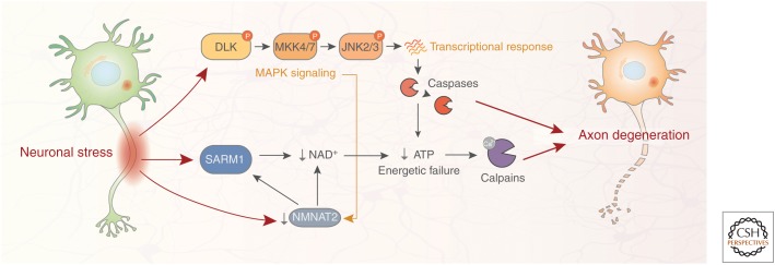
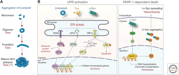
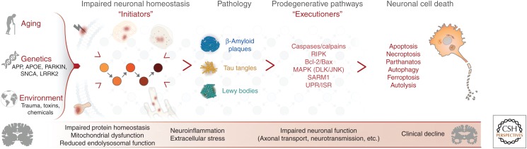
Neurodegenerative disease is characterized by the progressive deterioration of neuronal function caused by the degeneration of synapses, axons, and ultimately the death of nerve cells. An increased understanding of the mechanisms underlying altered cellular homeostasis and neurodegeneration is critical to the development of effective treatments for disease. Here, we review what is known about neuronal cell death and how it relates to our understanding of neurodegenerative disease pathology. First, we discuss prominent molecular signaling pathways that drive neuronal loss, and highlight the upstream cell biology underlying their activation. We then address how neuronal death may occur during disease in response to neuron intrinsic and extrinsic stressors. An improved understanding of the molecular mechanisms underlying neuronal dysfunction and cell death will open up avenues for clinical intervention in a field lacking disease-modifying treatments.


Figures

Figure 1.
Figure 1.
Apoptosis and necroptosis are executioners of neuronal cell death. (A) Neuronal apoptosis is executed by the caspase family of proteases. In the extrinsic pathway, death receptor engagement by secreted or membrane-bound death ligands leads to caspase-8 activation. In the intrinsic pathway, mitochondrial damage activates caspase-9 through back-mediated release of mitochondrial proteins. The intrinsic and extrinsic pathways converge on caspase-3 (and to a lesser extent in neurons, caspase-7), ultimately resulting in cell death. (B) Neuronal necroptosis is induced by several stimuli, including TNF signaling. Downstream from receptor activation, RIPK3 phosphorylates MLKL, leading to plasma membrane permeabilization and cell death. In addition to necroptosis, RIPK activity also mediates a cell-autonomous inflammation response that exacerbates neurodegeneration. TNF, Tumor necrosis factor; TNFR1, TNF receptor 1; RIPK, receptor-interacting kinase protein; MLKL, mixed lineage kinase domain-like protein; IL, interleukin.
Figure 2.
Figure 2.
Neuronal signaling pathways regulating axon degeneration. Axon degeneration is an active process involving several signaling pathways engaged by diverse neuronal stressors, both extracellular (e.g., axonal injury, toxins/chemotherapeutic agents) and intracellular (e.g., impaired protein homeostasis and mitochondrial dysfunction). Axonal damage results in both depletion of the protective axonal maintenance factor NMNAT2 and activation of SARM1, leading to a reduction of NAD+ levels, depletion of axonal ATP, and activation of proteolytic calpains. Neuronal stress also results in phosphorylation of DLK, a master kinase of the neuronal stress response that regulates both axon degeneration and neuronal apoptosis. DLK activation leads to phosphorylation of downstream kinases MKK4/7 and JNK2/3, and ultimately induces a prodegenerative caspase signaling cascade that activates calpains. There is cross talk between the SARM1 and DLK pathways; DLK signaling promotes NMNAT2 turnover, thereby enhancing SARM1-mediated axon degeneration. SARM1, sterile α and TIR motif-containing protein 1; NMNAT2, nicotinamide nucleotide adenylyltransferase 2; JNK, c-Jun amino-terminal kinase; DLK, dual leucine zipper kinase; MAPK, mitogen-activated protein kinase.
Figure 3.
Figure 3.
Impaired protein homeostasis is an initiator of neuronal cell death. (A) Aggregation of misfolded proteins, including β-amyloid, is a common pathological trait of chronic neurodegeneration disease. Monomeric proteins misfold and aggregate into toxic oligomers that recruit normal proteins into growing protofibrils, eventually forming mature plaques. Recent evidence suggests that oligomers are particularly toxic in disease, a topic of ongoing debate (Verma et al. 2015). (B, left) Protein aggregates cause endoplasmic reticulum (ER) stress and activate the UPR, inducing PERK, ATF6, and IRE1α signaling cascades. PERK activation leads to the phosphorylation of eIF2α and subsequent reduction of protein synthesis and expression of the transcription factor ATF4. ATF6 and IRE1α signaling lead to the expression of the transcription factors ATF6f and XBP1s, respectively. These transcription factors induce cellular homeostasis gene expression, but can also drive expression of proapoptotic genes under prolonged ER stress. JNK signaling is also activated downstream from IRE1α, promoting apoptosis. (B, right) In PD, α-syn fibrils active NOS, leading to PARP-1 activation and death of dopaminergic neurons via parthanatos. PAR produced by PARP-1 activation binds α-syn, increasing its aggregation, cell-to-cell spreading, and subsequent neurotoxicity. UPR, Unfolded protein response; PERK, PKR-like ER kinase; ATF4/6, activating transcription factor 4/6; IRE1α, inositol-requiring protein 1α; eIF2α, eukaryotic translation initiation factor 2α; XBP1, X-box binding protein 1; JNK, c-Jun amino-terminal kinase; NOS, nitric oxide synthase; NO, nitric oxide; PARP-1, poly(adenosine 5′-diphosphate-ribose) polymerase-1.
Figure 4.
Figure 4.
A model linking the initiators and executioners of neurodegenerative disease. A combination of aging, genetics, and environment contribute to the development of chronic neurodegenerative disease. These factors eventually lead to impaired neuronal homeostasis in response to neuron intrinsic and extrinsic disease initiators, including impaired protein homeostasis, mitochondrial dysfunction, neuroinflammation, among others. Over time, this altered homeostasis can manifest as pathologies commonly observed in disease, such as β-amyloid plaques and tau tangles in Alzheimer's disease (AD) or Lewy bodies in Parkinson's disease (PD). Neurons respond to these assaults by activating context-dependent, prodegenerative executioner pathways, precipitating impaired function and ultimately cell death. Disease-specific, stereotyped patterns of degeneration correlate with the clinical severity of neurodegenerative disease.

References

    1. Abisambra JF, Jinwal UK, Blair LJ, O'Leary JC III, Li Q, Brady S, Wang L, Guidi CE, Zhang B, Nordhues BA, et al. 2013. Tau accumulation activates the unfolded protein response by impairing endoplasmic reticulum-associated degradation. J Neurosci 33: 9498–9507. 10.1523/jneurosci.5397-12.2013 - DOI - PMC - PubMed
    1. Allan SM, Tyrrell PJ, Rothwell NJ. 2005. Interleukin-1 and neuronal injury. Nat Rev Immunol 5: 629–640. 10.1038/nri1664 - DOI - PubMed
    1. Arrazola MS, Saquel C, Catalan RJ, Barrientos SA, Hernandez DE, Catenaccio A, Court FA. 2019. Axonal degeneration is mediated by necroptosis activation. J Neurosci 39: 3832–3844. 10.1523/jneurosci.0881-18.2019 - DOI - PMC - PubMed
    1. Arriagada PV, Growdon JH, Hedley-Whyte ET, Hyman BT. 1992. Neurofibrillary tangles but not senile plaques parallel duration and severity of Alzheimer's disease. Neurology 42: 631–639. 10.1212/WNL.42.3.631 - DOI - PubMed
    1. Ashrafi G, Schlehe JS, LaVoie MJ, Schwarz TL. 2014. Mitophagy of damaged mitochondria occurs locally in distal neuronal axons and requires PINK1 and Parkin. J Cell Biol 206: 655–670. 10.1083/jcb.201401070 - DOI - PMC - PubMed