Review
. 2019 Dec 2;11(12):a032383.
doi: 10.1101/cshperspect.a032383.

Structural Biology of Telomerase

Affiliations
Review

Structural Biology of Telomerase

Yaqiang Wang et al. Cold Spring Harb Perspect Biol. .

Abstract

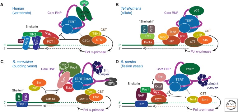
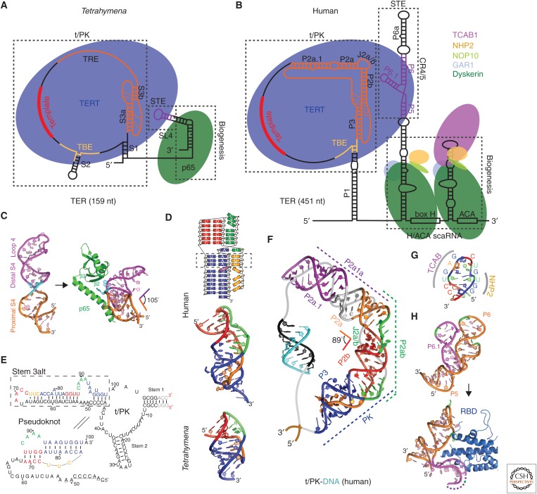
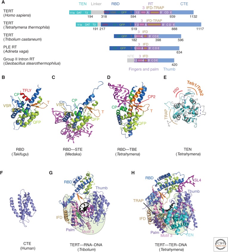
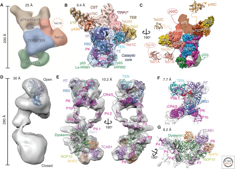
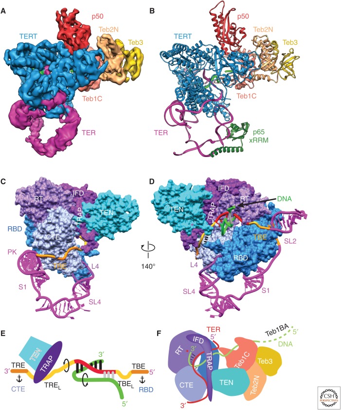
Telomerase is a DNA polymerase that extends the 3' ends of chromosomes by processively synthesizing multiple telomeric repeats. It is a unique ribonucleoprotein (RNP) containing a specialized telomerase reverse transcriptase (TERT) and telomerase RNA (TER) with its own template and other elements required with TERT for activity (catalytic core), as well as species-specific TER-binding proteins important for biogenesis and assembly (core RNP); other proteins bind telomerase transiently or constitutively to allow association of telomerase and other proteins with telomere ends for regulation of DNA synthesis. Here we describe how nuclear magnetic resonance (NMR) spectroscopy and X-ray crystallography of TER and protein domains helped define the structure and function of the core RNP, laying the groundwork for interpreting negative-stain and cryo electron microscopy (cryo-EM) density maps of Tetrahymena thermophila and human telomerase holoenzymes. As the resolution has improved from ∼30 Å to ∼5 Å, these studies have provided increasingly detailed information on telomerase architecture and mechanism.


Figures

Figure 1.
Figure 1.
Telomerase ribonucleoprotein (RNP) complexes and their interactions at telomeres. Telomerase RNPs and telomere-associated proteins and interactions are illustrated schematically for (A) human, (B) Tetrahymena thermophila, (C) budding yeast (Saccharomyces cerevisiae), and (D) fission yeast (Schizosaccharomyces pombe). Protein identities are indicated and colors chosen to highlight structural or functional conservation between species. Three principal complexes are illustrated: The telomerase core RNP (TER–TERT catalytic core and TER-binding proteins), telomerase accessory proteins (CST complexes, p50/TPP1/Est3, TEB/POT1), and shelterin/shelterin-like complexes. Black lines with arrows represent recruitment and/or enhancement of activity, whereas blunted black lines denote inhibition or termination of activity.
Figure 2.
Figure 2.
Tetrahymena and human telomerase core RNP and structures of TER domains. Schematics of telomerase core RNP of (A) Tetrahymena and (B) human telomerase. Proteins are shown at their approximate relative size and interactions, and the shape of TER is based on structural studies described in the text. (C) Nuclear magnetic resonance (NMR) structure of the free Tetrahymena stem-terminus element (STE; stem loop 4) (Protein Data Bank [PDB]: 2FEY) and model of the Tetrahymena p65 atypical RNA recognition motif (xRRM)–stem loop 4 complex based on the crystal structure of p65 xRRM–stem 4 (PDB: 4ERD) and NMR structure of loop 4 (PDB: 2M21). (D) Solution structures of minimal pseudoknots (PKs) from human (PDB: 2K95) and Tetrahymena (PDB: 5KMZ). Secondary structure schematic of the human P2b–P3 minimal PK is shown on the top. Dashed rectangle highlights the three U-A-U triples. (E) Comparison of secondary structures of free Tetrahymena TER and the folded PK, determined by NMR (Cash and Feigon 2017). The template residues are underlined. (F) Molecular model of human template/pseudoknot domain (t/PK). The solution NMR structure of the minimal PK (P2b/P3), P2b–J2a/b–P2a (P2ab), and residual dipolar coupling (RDC)–MC-Sym model of P2a.1–J2a.1–P2a (P2a1a) were computationally combined to model the full-length P2/P3 pseudoknot (Zhang et al. 2010). The single-stranded region (gray) containing the template (black) is shown bound to telomeric DNA (cyan). (G) Solution NMR structure of the 3′ apical stem loop (conserved region 7 [CR7]) containing the TCAB1 and NHP2 binding interfaces, determined by NMR and mutagenesis (Theimer et al. 2007). (H) NMR structure of free medaka (Oryzias latipes) STE (CR4/5) (PDB: 2MHI) compared with crystal structure of the CR4/5–RNA-binding domain (RBD) complex (PDB: 4O26).
Figure 3.
Figure 3.
TERT domains and structures. (A) Schematics comparing domains of reverse transcriptases (RTs) from human telomerase, Tetrahymena telomerase, Tribolium castaneum telomerase, Penelope-like element (PLE; Adineta vaga), and Group II intron (Geobacillus stearothermophilus). Domains and conserved motifs are aligned using the RT domains. Note that Tribolium “TERT” is more similar to the A. vaga PLE RT than to the true TERTs from human and Tetrahymena. Crystal structures (BG) of (B) Takifugu rubripes RBD (PDB: 4LMO), (C) medaka RBD–STE complex (PDB: 4O26), (D) Tetrahymena RBD–template boundary element (TBE) complex (PDB: 5C9H), and (E) free Tetrahymena telomerase amino-terminal domain (TEN) (PDB: 2B2A, cyan) superimposed with cryo-EM structure of TEN in Tetrahymena telomerase holoenzyme (PDB: 6D6V) (gray). Regions interacting with p50, TEB, and TRAP are indicated. Blue dashed lines represent the missing loops in the crystal structure. (F) Human carboxy-terminal element (CTE) (PDB: 5UGW) and (G) Tribolium TERT with an RNA–DNA hairpin mimicking a template–DNA duplex (PDB: 3KYL). (H) Cryo-EM structure of Tetrahymena TERT–TER with template–DNA duplex (PDB: 6D6V). TRAP is mostly covered by TEN, so is difficult to see in this view. The TRAP and TEN domains are unique to TERT, whereas motif 3 and IFD are found in closely related RTs from group II introns and PLEs. For G and H, the polymerase “hand” view is shown.
Figure 4.
Figure 4.
Comparison of negative-stain and cryo-EM density maps and models of Tetrahymena and human telomerase. (A–C) EM density maps and models of Tetrahymena telomerase. (A) A 25 Å negative-stain EM map with locations and estimated boundaries of subunits colored, based on affinity tagging of subunits, comparison of particles lacking all or part of a subunit, and modeling of the catalytic core (Jiang et al. 2013) (EMDB: 5804). (B) A 9.4 Å resolution cryo-EM map (core RNP, blue; CST, tan; TEB, straw; and p50, red) and pseudoatomic models of the core RNP and TEB and CST trimerization domains of three OB folds (Jiang et al. 2015). (C) An 180° rotated view of B with modeled domains shown as space-fill on the cryo-EM map. Model of the catalytic core and TEB is based on the 8.9 Å resolution map. Additional domains of Teb1, Teb2, p45, p65, and p50 are not visible in the cryo-EM map because of positional dynamics and are shown as crystal structures (Teb1A [PDB: 3U4V], Teb1B [PDB: 3U4Z], p45C [PDB: 5DFN]), homology models (Teb2C based on PDB: 1DPU), or ovals. (D–G) EM density maps and models of human telomerase. (D) A 30 Å negative-stain EM map originally proposed to be a dimer (EMDB: 2310) (Sauerwald et al. 2013). Tribolium TERT was automatically fit into the map using UCSF Chimera (Pettersen et al. 2004). (E) Two views of the 10.2 Å resolution cryo-EM map of human telomerase holoenzyme (EMDB: 7521) with a modeled catalytic core (top) and H/ACA scaRNP (bottom) (Nguyen et al. 2018). (F) A 7.7 Å resolution cryo-EM map (EMDB: 7518) from focused refinement and model of the catalytic core. (G) A 8.2 Å resolution map (EMDB: 7519) from focused refinement and model of the H/ACA scaRNP. TER is magenta in all models and TERT is blue. Proteins and TER domains are labeled.
Figure 5.
Figure 5.
Cryo-EM of Tetrahymena telomerase at 4.8 Å resolution provides insights into the mechanism. (A) A 4.8 Å resolution cryo-EM density map of Tetrahymena telomerase with telomeric DNA. (B) Atomic model of the TERT–TER catalytic core, p65 xRRM, p50, and TEB. The model of p65 xRRM is based on the crystal structure (PDB: 4ERD). (C-D) Two views of the structure of the telomerase catalytic core with TERT as spacefill and TER as ribbon. TERT and TER domains are labeled. TEN and TRAP are connected to the RBD and RT domains, respectively, from opposite sides of the TERT ring, physically interlocking the t/PK and TERT. In D, black dashes between the TEN and RBD domains denote the linker between them. The nascent telomeric DNA is shown exiting the template. (E) Cartoon of template recognition element (TRE)–template–TBE interactions with TERT and telomeric DNA. Arrows indicate directions of rotation of TER and DNA during telomere repeat synthesis, and anchors correspond to TER anchor sites that define the template boundaries. TRE linker (TREL) and the TBE loop (TBEL) are the single-stranded regions between the template and TRE and TBE, respectively. (F) Cartoon illustrating the paths of TRE–template–TBE on TERT and of telomeric DNA from the TERT active site to TEB; the TRAP–TEN interaction; the TRAP–TRE interaction. The protein p50 is omitted from this schematic for clarity. (Figure panels are modified from Jiang et al. 2018.)

Similar articles

Cited by

References

    1. Adams PD, Afonine PV, Bunkoczi G, Chen VB, Davis IW, Echols N, Headd JJ, Hung LW, Kapral GJ, Grosse-Kunstleve RW, et al. 2010. PHENIX: A comprehensive Python-based system for macromolecular structure solution. Acta Crystallogr D Biol Crystallogr 66: 213–221. - PMC - PubMed
    1. Akiyama BM, Stone MD. 2013. Structural biology: A solution to the telomerase puzzle. Nature 496: 177–178. - PubMed
    1. Akiyama BM, Loper J, Najarro K, Stone MD. 2012. The C-terminal domain of Tetrahymena thermophila telomerase holoenzyme protein p65 induces multiple structural changes in telomerase RNA. RNA 18: 653–660. - PMC - PubMed
    1. Akiyama BM, Parks JW, Stone MD. 2015. The telomerase essential N-terminal domain promotes DNA synthesis by stabilizing short RNA-DNA hybrids. Nucleic Acids Res 43: 5537–5549. - PMC - PubMed
    1. Angrisani A, Vicidomini R, Turano M, Furia M. 2014. Human dyskerin: Beyond telomeres. Biol Chem 395: 593–610. - PubMed

Publication types

MeSH terms