. 2019 Oct 7;9(10):3489-3500.
doi: 10.1534/g3.119.400554.

Destabilization of the Tumor-Inducing Plasmid from an Octopine-Type Agrobacterium tumefaciens Lineage Drives a Large Deletion in the Co-resident At Megaplasmid

Affiliations

Destabilization of the Tumor-Inducing Plasmid from an Octopine-Type Agrobacterium tumefaciens Lineage Drives a Large Deletion in the Co-resident At Megaplasmid

Ian S Barton et al. G3 (Bethesda). .

Abstract

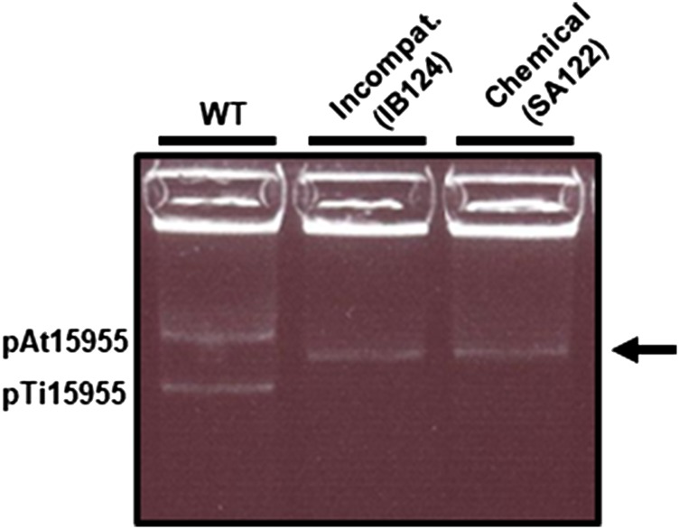
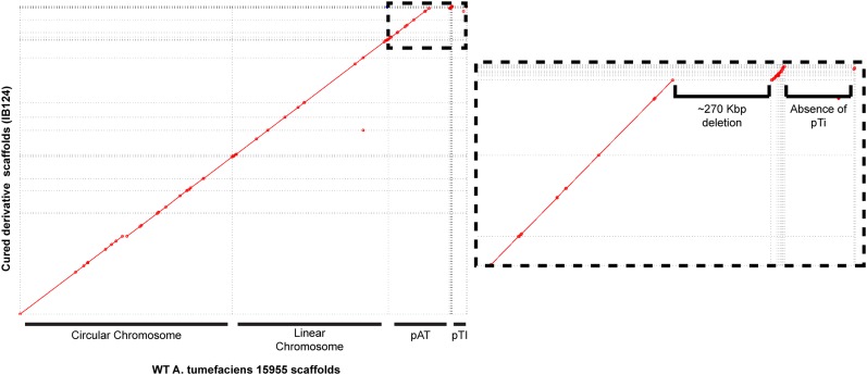
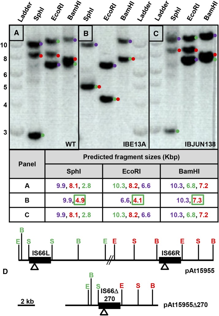
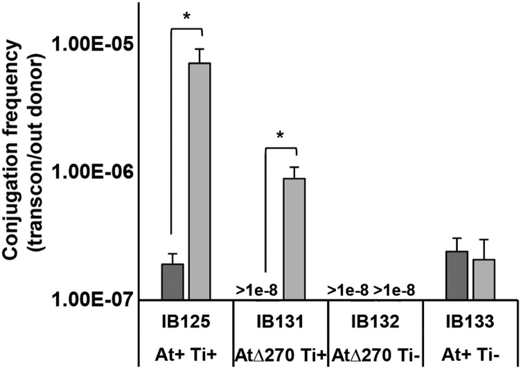
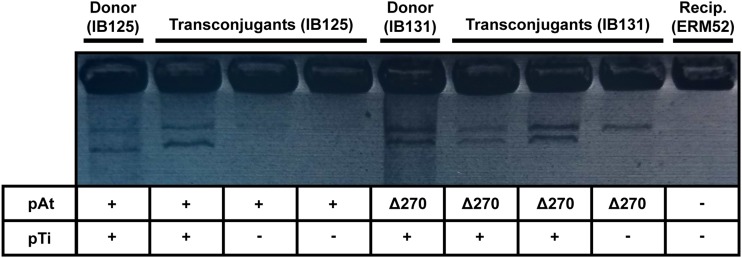
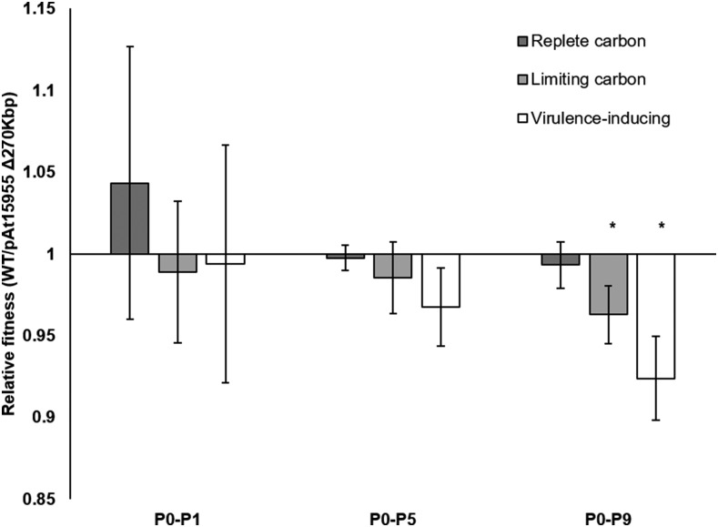
Bacteria with multi-replicon genome organizations, including members of the family Rhizobiaceae, often carry a variety of niche-associated functions on large plasmids. While evidence exists for cross-replicon interactions and co-evolution between replicons in many of these systems, remarkable strain-to-strain variation is also observed for extrachromosomal elements, suggesting increased genetic plasticity. Here, we show that curing of the tumor-inducing virulence plasmid (pTi) of an octopine-type Agrobacterium tumefaciens lineage leads to a large deletion in the co-resident At megaplasmid (pAt). The deletion event is mediated by a repetitive IS-element, IS66, and results in a variety of environment-dependent fitness consequences, including loss of independent conjugal transfer of the plasmid. Interestingly, a related and otherwise wild-type A. tumefaciens strain is missing exactly the same large pAt segment as the pAt deletion derivatives, suggesting a similar event over its natural history. Overall, the findings presented here uncover a novel genetic interaction between the two large plasmids of A. tumefaciens and provide evidence for cross-replicon integration and co-evolution of these plasmids.

Keywords: IS element; Ti plasmid; accessory genome; conjugal transfer; deletion; plasmid curing.


Figures

Figure 1
Figure 1
Multiple changes in plasmid profile of A. tumefaciens 15955 upon curing of pTi15955. Eckhardt gel electrophoresis analysis of WT A. tumefaciens 15955 and two derivatives of A. tumefaciens 15955, cured of the Ti plasmid (pTi15955) using either chemical (SA122) or plasmid incompatibility methods (IB124). Positions of pAt15955 and pTi15955 in WT A. tumefaciens 15955 are shown. Arrow indicates single band present in pTi15955-cured derivatives.
Figure 2
Figure 2
Whole genome sequencing reveals co-incident pAt15955 deletion upon pTi15955 curing. Mummerplot visualization of MUMmer 3 alignment of genome assembly scaffolds from WT 15955 (X axis) and a pTi15955-cured derivative (IB124, Y axis). Matching sequence between assemblies is indicated with diagonal red line segments. Sequence corresponding to each replicon is indicated. Inset (dashed) highlights large (270 Kbp) deletion of pAt15955 and absence of pTi15955 in IB124.
Figure 3
Figure 3
Loss of one IS66 element upon deletion of ∼270 Kbp pAt15955 segment. Southern blot analysis of gDNA from WT A. tumefaciens 15955 (A) and two derivatives that have either incurred loss of the pAt15955 segment (IBE13A, B) or pTi15955 (IBJUN138, C) using three restriction enzyme digests that do not cleave within IS66 (SphI, EcoRI, and BamHI). Ladder shown is NEB 1 kb DNA ladder. Numbers on Y-axis indicated ladder sizes in Kbp. Prediction of IS66-containing restriction fragment sizes for digests in A, B, and C – color coding matches the dots adjacent to each band; Blue, IS66 on the linear chromosome; Green, IS66L; Red IS66R; New restriction fragments generated by the deletion are red text boxed green (D) Restriction site locations in the intact pAt15955 plasmid and the pAt15955Δ270 derivative. S, SphI; E, EcoRI, B, BamHI. Color coding of restriction sites matches predicted fragments and the annotated blot image. Open triangles represent the location of the probe fragment used for the hybridizations, and the double hash marks are the remaining 270 kb deleted from pAt15955.
Figure 4
Figure 4
Octopine-inducible pAt15955 conjugation is pTi15955-dependent and independent of pAt15955-encoded trb machinery. pAt15955 conjugation frequencies of marked (KmR) derivatives of A. tumefaciens 15955 (donors) into a plasmidless recipient, ERM52, under standard growth (ATGN, dark gray) and octopine-containing (ATGNO, hatched, 3.25 mM) conditions. Donor strains shown are WT (IB125) or derivatives deleted for pAt15955 segment (IB131), pTi15955 (IB133), or both (IB132). Conjugation frequencies reported as transconjugants per output donor (see Materials and Methods). The * denotes a p-value <0.03.
Figure 5
Figure 5
Co-transfer of pTi15955 in pAt15955 transconjugants in the presence of octopine. Plasmid profiles for subset of pAt15955 transconjugants from conjugation assay in Figure 4 were determined by Eckhardt gel electrophoresis. Shown are profiles for two donors (IB125 and IB131), recipient (ERM52), and three transconjugants from conjugation assays with either donor (IB125 or IB131). Indicated below gel image are presence (+) or absence (-) of pTi15955 and the presence of WT pAt15955 (+) or pAt15955∆270 (∆270).
Figure 6
Figure 6
Absence of pAt15955 segment confers an environment-dependent fitness advantage. Relative fitness of WT A. tumefaciens 15955 and a strain isogenic for loss of the pAt15955 segment over one (P0-P1), five (P0-P5) and nine (P0-P9) serial passages in replete carbon (dark gray), limiting carbon (hatched), and virulence-inducing conditions (white). Bars represent 6 biological replicates, 3 each with inversely marked strains (Methods). Relative fitness calculated as the total number of doublings of each competitor over the indicated passing interval (Methods). Error bars shown are standard deviation. Asterisk indicates p-value < 0.05 in comparison to the expected value of 1.

References

    1. Agnoli K., Schwager S., Uehlinger S., Vergunst A., Viteri D. F. et al. , 2012. Exposing the third chromosome of Burkholderia cepacia complex strains as a virulence plasmid. Mol. Microbiol. 83: 362–378. 10.1111/j.1365-2958.2011.07937.x - DOI - PubMed
    1. Bailey J., and Manoil C., 2002. Genome-wide internal tagging of bacterial exported proteins. Nat. Biotechnol. 20: 839–842. 10.1038/nbt715 - DOI - PubMed
    1. Barnett M. J., Toman C. J., Fisher R. F., and Long S. R., 2004. A dual-genome Symbiosis Chip for coordinate study of signal exchange and development in a prokaryote–host interaction. Proc. Natl. Acad. Sci. USA 101: 16636–16641. 10.1073/pnas.0407269101 - DOI - PMC - PubMed
    1. Bell J. C., and Kowalczykowski S. C., 2016. RecA: Regulation and mechanism of a molecular search engine. Trends Biochem. Sci. 41: 491–507. Erratum 41: 646. 10.1016/j.tibs.2016.04.002 - DOI - PMC - PubMed
    1. Bellanger X., Payot S., Leblond-Bourget N., and Guedon G., 2014. Conjugative and mobilizable genomic islands in bacteria: evolution and diversity. FEMS Microbiol. Rev. 38: 720–760. 10.1111/1574-6976.12058 - DOI - PubMed

Publication types

LinkOut - more resources