. 2019 Oct 18;87(11):e00512-19.
doi: 10.1128/IAI.00512-19. Print 2019 Nov.

Identification of Novel Bacteriophages with Therapeutic Potential That Target Enterococcus faecalis

Affiliations

Identification of Novel Bacteriophages with Therapeutic Potential That Target Enterococcus faecalis

M Al-Zubidi et al. Infect Immun. .

Abstract

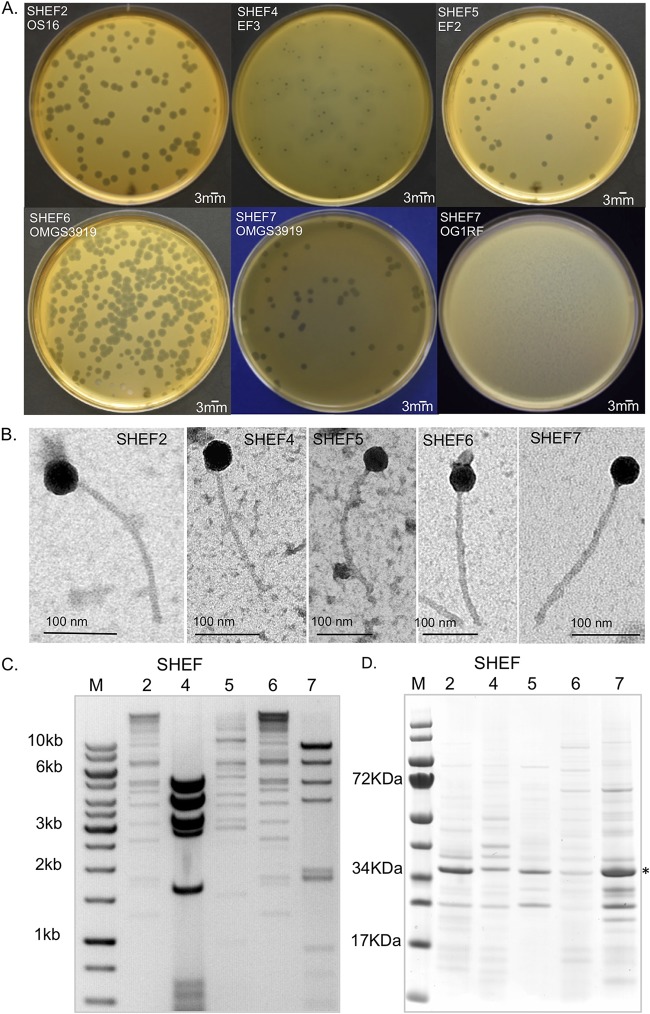
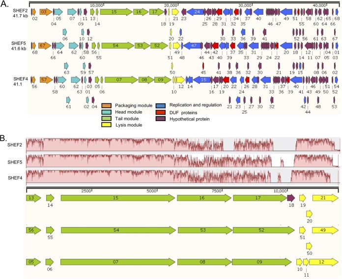
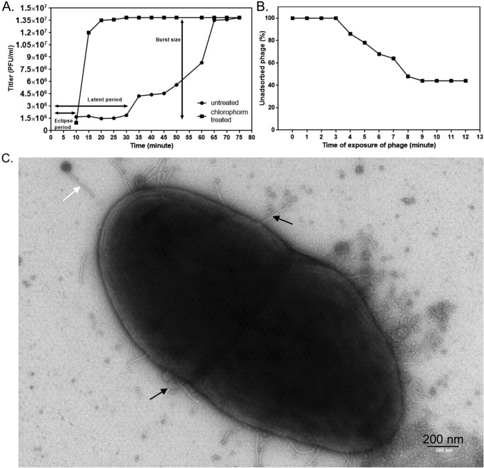
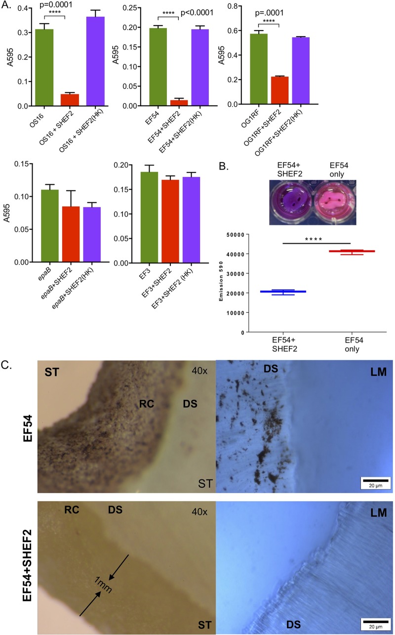
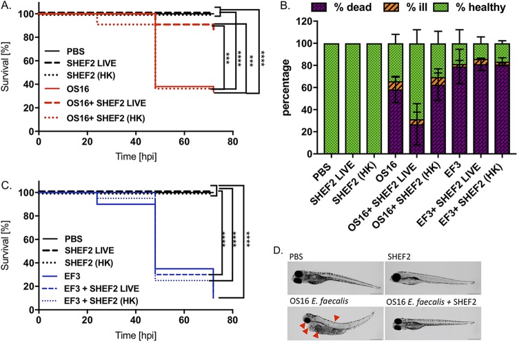
The Gram-positive opportunistic pathogen Enterococcus faecalis is frequently responsible for nosocomial infections in humans and represents one of the most common bacteria isolated from recalcitrant endodontic (root canal) infections. E. faecalis is intrinsically resistant to several antibiotics routinely used in clinical settings (such as cephalosporins and aminoglycosides) and can acquire resistance to vancomycin (vancomycin-resistant enterococci). The resistance of E. faecalis to several classes of antibiotics and its capacity to form biofilms cause serious therapeutic problems. Here, we report the isolation of several bacteriophages that target E. faecalis strains isolated from the oral cavity of patients suffering root canal infections. All phages isolated were Siphoviridae with similar tail lengths (200 to 250 nm) and icosahedral heads. The genome sequences of three isolated phages were highly conserved with the exception of predicted tail protein genes that diverge in sequence, potentially reflecting the host range. The properties of the phage with the broadest host range (SHEF2) were further characterized. We show that this phage requires interaction with components of the major and variant region enterococcal polysaccharide antigen to engage in lytic infection. Finally, we explored the therapeutic potential of this phage and show that it can eradicate E. faecalis biofilms formed in vitro on a standard polystyrene surface but also on a cross-sectional tooth slice model of endodontic infection. We also show that SHEF2 cleared a lethal infection of zebrafish when applied in the circulation. We therefore propose that the phage described here could be used to treat a broad range of antibiotic-resistant E. faecalis infections.

Keywords: bacteriophage; biofilm; capsule; oral microbiology.


Figures

FIG 1
FIG 1
Isolation of E. faecalis bacteriophage and plaque morphology. (A) Images of bacterial plaques formed by the isolated phage in top-agar lawns of E. faecalis OS16 (SHEF2), E. faecalis EF3 (SHEF4), E. faecalis EF2 (SHEF5), and E. faecalis OMGS3919 (SHEF6 and -7). SHEF7 is also shown with E. faecalis OG1RF to illustrate plaque morphologies. (B) Transmission electron micrographs of SHEF phage particles. Phages were negatively stained with 0.2% uranyl acetate as described in Materials and Methods. Scale bars, 100 nm. (C) RFLP analysis of extracted phage chromosomal DNA. SHEF2, -4, -5, and -7 phage genomic DNA was digested with HindIII and analyzed by agarose gel electrophoresis (an inverted image is shown alongside the GeneRuler 1-kb ladder). (D) Virion protein profiles of SHEF2, -4, -5, -6, and -7 by SDS-PAGE with InstantBlue staining. The dominant protein band identified by MS/MS as the major capsid protein for SHEF2 at 36 kDa is indicated by an asterisk (*).
FIG 2
FIG 2
Genome organization of E. faecalis lytic phages SHEF2, SHEF5, and SHEF4. (A) Images produced using SnapGene Viewer 1.1.3 software (DUF, conserved domains of unknown function). Genome annotation corresponds to GenBank accession numbers MF678788, MF678789, and MF678790 for SHEF2, -4, and -5, respectively. The colors correspond to the predicted function, as indicated in the key. (B) Mauve alignment of tail and lysis genes highlighting areas of conservation.
FIG 3
FIG 3
Molecular determination of SHEF2 phage adhesion using strain OG1RF. (A) Schematic of the OG1RF epa core (purple) and variable locus (blue), generated using SnapGene (labeling is according to accession number NC_017316.1). (B) Table showing qualitative results of spot assay double-layer agar infections of strains listed with SHEF2 (+, infection; –, no infection) (for pictures, see Fig. S4). (C) Phage adsorption assay for OG1RF and its isogenic epaB and OPDV_11720 mutant strains (all at 108 CFU/ml bacteria) with SHEF2. Phages (input, 2 × 106) were added for 24 h before the phages were enumerated in cell supernatants before and after treatment with 0.28 M NaCl using a titer assay. ND, not determined (due to all cells being dead). This experiment was repeated twice, in triplicate each time (the mean is shown), with one example displayed here. (D) TEM images of infected OG1RF, epaB, and OPDV_11720 mutants with SHEF2 at 30 min postinfection. Arrows indicate adsorbed phage.
FIG 4
FIG 4
One-step growth and adsorption rate characterization of SHEF2. (A) One-step growth curve of SHEF2 phage with E. faecalis OS16 as the host. The two sets of data represent samples treated with or without chloroform. The eclipse, burst, and latent periods are labeled. (B) Adsorption of SHEF2 phage to E. faecalis OS16 expressed as a percentage of the total phages added. (C) Transmission electron micrograph of strain OS16 + SHEF2 at 30 min postinfection. Black arrows, spent heads and adsorbed phage; white arrow, unadsorbed phage.
FIG 5
FIG 5
Biofilm assay on polystyrene plates and tooth root slices. (A) Bar charts represent biofilm growth, as measured by crystal violet staining (measured using the A570 of the extracted stain, normalized to the cell growth in each well [A600]). Samples treated with live phage are labeled “+SHEF2,” while those with heat-killed [+SHEF2 (HK)] strain names are as shown elsewhere. Means of six polystyrene microtiter wells per condition are shown along with the SD, and a Student t test was used to compare conditions (P < 0.0001). Experiments were conducted on three separate occasions; one example is shown here. (B, upper panel) Photograph of tooth root slices in situ treated with SHEF2 (+SHEF2) or with strain EF54 only. The difference in color represents the resazurin reduction in response to resorufin and is represented quantitatively below (mean, SD from three readings). A Student t test was used to compare treated and untreated groups (P < 0.0001). (C) Stereomicroscope (ST, left) and light microscope (LM, right [resazurin stained]) images represent the untreated group (upper), while the lower images show SHEF2-treated samples. Biofilms of E. faecalis colonies scattered on the root canal surface (RC) and the dentinal surface (DS) of the ST and LM images, respectively, are shown.
FIG 6
FIG 6
Phage SHEF2 treatment of E. faecalis OS16-infected zebrafish embryos. Zebrafish were infected systematically with E. faecalis OS16 strain or strain EF3 at a dose of 30,000 CFU at 30 h postfertilization. After 2 h, the embryos were injected with SHEF2 (SHEF2 LIVE) phage or heat-inactivated [SHEF2(HK)] at an MOI of 20 (in 2 nl). Control experiments were also performed with phage only [SHEF2 LIVE or SHEF2(HK)] and PBS (2 nl). Data are presented as Kaplan-Meier survival plots (A and C), as well as a bar chart (B), indicating mortality data at 72 hpi or all conditions. Bars represent means ± the SD. Three independent experiments were performed (n = 20 zebrafish per condition per experiment). Statistical comparison between groups was performed using a log-rank test (A and C) or one-way ANOVA (B). (D) Morphology of zebrafish embryos at 72 hpi after injection with PBS, E. faecalis OS16 alone, SHEF2 LIVE alone, or E. faecalis OS16, followed by SHEF2 LIVE (D). Red arrows indicate symptoms of infection (eye and yolk sac abnormalities, spinal curving, and pericardial edema). Scale bar, 500 μm.

Similar articles

Cited by

References

    1. Wisplinghoff H, Bischoff T, Tallent SM, Seifert H, Wenzel RP, Edmond MB. 2004. Nosocomial bloodstream infections in US hospitals: analysis of 24,179 cases from a prospective nationwide surveillance study. Clin Infect Dis 39:309–317. doi:10.1086/421946. - DOI - PubMed
    1. Gilmore MS, Lebreton F, van Schaik W. 2013. Genomic transition of enterococci from gut commensals to leading causes of multidrug-resistant hospital infection in the antibiotic era. Curr Opin Microbiol 16:10–16. doi:10.1016/j.mib.2013.01.006. - DOI - PMC - PubMed
    1. Paulsen IT, Banerjei L, Myers GSA, Nelson KE, Seshadri R, Read TD, Fouts DE, Eisen JA, Gill SR, Heidelberg JF, Tettelin H, Dodson RJ, Umayam L, Brinkac L, Beanan M, Daugherty S, DeBoy RT, Durkin S, Kolonay J, Madupu R, Nelson W, Vamathevan J, Tran B, Upton J, Hansen T, Shetty J, Khouri H, Utterback T, Radune D, Ketchum KA, Dougherty BA, Fraser CM. 2003. Role of mobile DNA in the evolution of vancomycin-resistant Enterococcus faecalis. Science 299:2071–2074. doi:10.1126/science.1080613. - DOI - PubMed
    1. Polidori M, Nuccorini A, Tascini C, Gemignani G, Iapoce R, Leonildi A, Tagliaferri E, Menichetti F. 2011. Vancomycin-resistant Enterococcus faecium (VRE) bacteremia in infective endocarditis successfully treated with combination daptomycin and tigecycline. J Chemother 23:240. doi:10.1179/joc.2011.23.4.240. - DOI - PubMed
    1. Love R. 2001. Enterococcus faecalis–a mechanism for its role in endodontic failure. Int Endod J 34:399–405. doi:10.1046/j.1365-2591.2001.00437.x. - DOI - PubMed

Publication types