. 2019 Sep 10;116(37):18435-18444.
doi: 10.1073/pnas.1910574116. Epub 2019 Aug 26.

Chemoptogenetic damage to mitochondria causes rapid telomere dysfunction

Affiliations

Chemoptogenetic damage to mitochondria causes rapid telomere dysfunction

Wei Qian et al. Proc Natl Acad Sci U S A. .

Erratum in

Abstract

Reactive oxygen species (ROS) play important roles in aging, inflammation, and cancer. Mitochondria are an important source of ROS; however, the spatiotemporal ROS events underlying oxidative cellular damage from dysfunctional mitochondria remain unresolved. To this end, we have developed and validated a chemoptogenetic approach that uses a mitochondrially targeted fluorogen-activating peptide (Mito-FAP) to deliver a photosensitizer MG-2I dye exclusively to this organelle. Light-mediated activation (660 nm) of the Mito-FAP-MG-2I complex led to a rapid loss of mitochondrial respiration, decreased electron transport chain complex activity, and mitochondrial fragmentation. Importantly, one round of singlet oxygen produced a persistent secondary wave of mitochondrial superoxide and hydrogen peroxide lasting for over 48 h after the initial insult. By following ROS intermediates, we were able to detect hydrogen peroxide in the nucleus through ratiometric analysis of the oxidation of nuclear cysteine residues. Despite mitochondrial DNA (mtDNA) damage and nuclear oxidative stress induced by dysfunctional mitochondria, there was a lack of gross nuclear DNA strand breaks and apoptosis. Targeted telomere analysis revealed fragile telomeres and telomere loss as well as 53BP1-positive telomere dysfunction-induced foci (TIFs), indicating that DNA double-strand breaks occurred exclusively in telomeres as a direct consequence of mitochondrial dysfunction. These telomere defects activated ataxia-telangiectasia mutated (ATM)-mediated DNA damage repair signaling. Furthermore, ATM inhibition exacerbated the Mito-FAP-induced mitochondrial dysfunction and sensitized cells to apoptotic cell death. This profound sensitivity of telomeres through hydrogen peroxide induced by dysregulated mitochondria reveals a crucial mechanism of telomere-mitochondria communication underlying the pathophysiological role of mitochondrial ROS in human diseases.

Keywords: ATM signaling; DNA damage response; mitochondria; singlet oxygen; telomere.


Conflict of interest statement

Conflict of interest statement: M.P.B. is a founder of Sharp Edge Labs, a company applying the FAP-fluorogen technology.

Figures
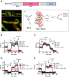
Fig. 1.
Fig. 1.
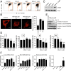
ROS exclusively generated in mitochondria by a Mito-FAP system induces mitochondrial dysfunction. (A) The structure of the Mito-FAP plasmid construct (i), STED images of localization of Mito-FAP (ii and iii), and the mechanism of generation of singlet oxygen on binding of MG-2I with FAP after light exposure (iv). A, ii shows a low-magnification single-plane STED image of HEK293 cells expressing mNEON-TOM20 (green) and Mito-FAP (red; imaged through binding to MG-ester). The red signal of Mito-FAP is clearly contained within the green mNEON profile. This is more clearly shown in the enlargement (A, iii). (B and C) HEK293 cells stably expressing Mito-FAP were treated with MG-2I dye (50 nM) alone, light exposure (660 nm, 5 min) alone, or light exposure (660 nm, 5 min) in the presence of 50 nM MG-2I. Mitochondrial respiration as determined by OCR and ECAR was assessed by a Seahorse Extracellular Flux Analyzer 4 h after treatment. (D and E) HEK293 Mito-FAP cells were treated with or without a singlet oxygen scavenger sodium azide (50 mM; D) or a broad-spectrum ROS scavenger NAC (10 mM; E) added 15 min before exposure to MG-2I dye (50 nM) and light (5 min). Four hours after treatment, OCR was determined by a Seahorse Extracellular Flux Analyzer. Data show the representative results of at least 3 experiments with similar results. Data are represented as mean ± SD of at least 6 wells. MLS, mitochondrial leading sequence.
Fig. 2.
Fig. 2.
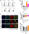
Characterization of mitochondrial damage induced by mitochondrial singlet oxygen generation through Mito-FAP. (A) HEK293 Mito-FAP cells were treated with MG-2I dye (50 nM) alone, light exposure (660 nm, 5 min) alone, or light exposure (660 nm, 5 min) in the presence of 50 nM MG-2I. The mitochondrial membrane potential was assessed by a membrane potential indicator JC-1 24 h after treatment. The loss of JC-1 red fluorescence after MG-2I and light treatment indicates the shift from JC-1 dimer to JC-1 monomer and hence, the loss of mitochondrial membrane potential. (B) At 4 and 24 h after treatment with 50 nM MG-2I and light (660 nm, 5 min), mitochondrial morphology was visualized by staining cells with Tom20 and Alexa 555-conjugated secondary antibody. Mitochondrial connectivity was quantified by reconstructed 3-dimensional images using Imaris software. (Scale bars: 5 μm.) (C) Phosphorylation of Drp1 was determined by western blot 24 h after treatment as indicated. (D) HEK293 Mito-FAP cells were treated with or without the combination of MG-2I (50 nM) and light (5 min). For the determination of ETC complex activities, cells were harvested right after treatment (0 h) or after 4 and 24 h of recovery. Data represent mean ± SEM of at least 3 independent experiments. *P < 0.05; **P < 0.01; ***P < 0.001. (E) Changes in the protein levels of several subunits of ETC complexes were evaluated by western blot at the indicated time points after MG-2I (50 nM) and light (5-min) treatment. (F) HEK293 Mito-FAP cells were treated with MG-2I dye (50 nM) alone, light exposure (660 nm, 5 min) alone, or light exposure (660 nm, 5 min) in the presence of 50 nM MG-2I. The damage of mtDNA and nuclear DNA was analyzed by a long-range qPCR DNA lesion assay 24 h after treatment. The PCR amplification of a large mtDNA segment (8.9 kb), a small mtDNA segment (221 bp), and the gene of polymerase β (12.2 kb) was performed. The inclusion of the measurement of amplification with a template DNA amount that is half of the amount used in control group indicates that the amount of PCR product corresponds to the starting amount of template DNA. Data represent mean ± SEM of 3 independent experiments with 3 PCR reactions per treatment condition. *P < 0.05 (one-way ANOVA); **P < 0.01 (one-way ANOVA).
Fig. 3.
Fig. 3.
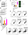
Singlet oxygen-induced mitochondrial dysfunction leads to a secondary wave generation of ROS. (A) Mitochondrial generation of ROS was determined by MitoSox at 4, 24, and 48 h after treatment of HEK293 Mito-FAP cells with MG-2I (50 nM), light (5 min), or light (5 min) in the presence of MG-2I (50 nM). (B) MitoTEMPO (100 nM) inhibited mitochondrial superoxide generation 4 h after treatment. ***P < 0.001. (C) Oxidation of protein SH group was evaluated by thiol oxidation immunocytochemistry 24 h after treatment with light (5 min) in the presence of MG-2I (50 nM). Treatment of 100 μM H2O2 for 20 min served as the positive control. Representative images are shown. (D) Quantification on the ratio of fluorescence intensity of Alexa Fluor 555/647 (SS/SH) in the nuclear region of the cells. Data are represented as mean ± SD. ***P < 0.001. (E) Detection of diffusion of H2O2 into nuclei by a fluorescent nuclear H2O2 sensor pHyper-nuc 24 h after treatment with light (5 min) in the presence of MG-2I (50 nM). Data are represented as mean ± SEM. *P < 0.05.
Fig. 4.
Fig. 4.
Mitochondrial dysfunction leads to nuclear DNA replication stress. (A) HEK293 Mito-FAP cells were treated with MG-2I (50 nM), light (5 min), or light (5 min) in the presence of MG-2I (50 nM). Cell proliferation was determined by a CyQuant assay at indicated time points. Data are represented as mean ± SD of 4 wells. A representative experiment is shown. (B) Apoptotic cell death in HEK293 Mito-FAP cells after treatment with MG-2I (50 nM), light (5 min), or light (5 min) in the presence of MG-2I (50 nM) was determined by Annexin V and PI staining at indicated time points. Numbers indicate the percentages of each population positive or negative with Annexin V or PI staining. (C) The effect of the exposure to light (5 min) in the presence of MG-2I (50 nM) on cell cycle progression was determined by BrdU incorporation (DNA replication) and phosphorylation of histone H3 (mitosis). Numbers indicate the percentages of cell population positive for incorporating BrdU or phosphorylated histone H3. (D) The phosphorylation of RPA32 and the protein level of Cyclin E after HEK293 Mito-FAP cells treated with light (5 min) in the presence of MG-2I (50 nM) were determined by western blot.
Fig. 5.
Fig. 5.
Activation of the ATM pathway in response to oxidative stress after MG-2I and light treatment. (A) At 24 h after MG-2I (50 nM) and light (5-min) treatment, phosphorylation of ATM and Chk2 was determined by western blot. (B) Cells were treated with NAC (10 mM) for 15 min before, during, and after exposure to MG-2I dye (50 nM) and light (5 min). At 24 h postexposure, the phosphorylation of ATM and Chk2 was determined by western blot. (C) DNA single-strand breaks were determined by alkali Comet assay 24 h after treatment with light (5 min) in the presence of MG-2I (50 nM). Treatment of 100 μM H2O2 for 20 min served as a positive control. Data are represented as mean ± SEM. Control (n = 235), MG-2I + Light (n = 263), and H2O2 (n = 91). ns, not significant. ****P < 0.0001. (D) Cells were treated with ATM kinase inhibitor KU-55933 alone, MG-2I (50 nM) plus light (5 min), or MG-2I plus light exposure in the presence of KU-55933. The generation of mitochondrial superoxide was determined 4 h after treatment by MitoSox. Data are represented as mean ± SEM. *P < 0.05. (E) Cells were treated in a manner as described in D, and the apoptosis was determined 72 h after MG-2I and light treatment. (F) The number of cells in mitosis was measured by the phosphorylation of histone H3 24 h after treatment.
Fig. 6.
Fig. 6.
Mitochondrial dysfunction leads to telomere damage. (A) The recruitment of 53BP1 (red) at the telomeres (green) 48 h after MG-2I (50 nM) and light (5-min) treatment was analyzed by immunofluorescence. The number of 53BP1+/PNA+ TIFs was quantified. Data are represented as mean ± SD. ****P < 0.0001. (Scale bars: 2 μm.) (B) The number of HEK293 Mito-FAP cells with fragile telomere and telomere loss (signal-free ends) was evaluated by FISH 48 h after MG-2I (50 nM) and light (5-min) treatment. Data are represented as mean ± SD. (Scale bar: 2 μm.) (C) Telomere damage in HEK293 Mito-FAP ρ0 cells was analyzed as described in B. Data are represented as mean ± SD. (D) HEK293 Mito-FAP cells were pretreated with NAC (10 mM) for 15 min before, during and after light (5-min) exposure, and their telomere damage was then analyzed as in B. Data are represented as mean ± SD. At least 50 cells were counted. Representative results from 3 independent experiments are shown. *P < 0.05, **P < 0.01, ***P < 0.001.
Fig. 7.
Fig. 7.
Working model of how mitochondrially generated hydrogen peroxide causes telomere damage. On 660-nm light exposure, the complex of Mito-FAP and MG-2I produces singlet oxygen. Singlet oxygen can induce oxidative damage to mitochondrial ETC, initiating a persistent secondary wave of superoxide and hydrogen peroxide generation. Hydrogen peroxide generated by mitochondria is able to damage mtDNA, which amplifies the damage to ETC. Hydrogen peroxide can further diffuse to the nucleus and is sufficient to cause nuclear protein oxidation and preferential telomere DNA damage but not overall nuclear DNA damage.

Similar articles

Cited by

References

    1. Schieber M., Chandel N. S., ROS function in redox signaling and oxidative stress. Curr. Biol. 24, R453–R462 (2014). - PMC - PubMed
    1. Zorov D. B., Juhaszova M., Sollott S. J., Mitochondrial reactive oxygen species (ROS) and ROS-induced ROS release. Physiol. Rev. 94, 909–950 (2014). - PMC - PubMed
    1. Van Houten B., Woshner V., Santos J. H., Role of mitochondrial DNA in toxic responses to oxidative stress. DNA Repair (Amst.) 5, 145–152 (2006). - PubMed
    1. de Moura M. B., dos Santos L. S., Van Houten B., Mitochondrial dysfunction in neurodegenerative diseases and cancer. Environ. Mol. Mutagen. 51, 391–405 (2010). - PubMed
    1. Yakes F. M., Van Houten B., Mitochondrial DNA damage is more extensive and persists longer than nuclear DNA damage in human cells following oxidative stress. Proc. Natl. Acad. Sci. U.S.A. 94, 514–519 (1997). - PMC - PubMed

Publication types

MeSH terms