. 2019 Aug 26;10(1):3854.
doi: 10.1038/s41467-019-11486-1.

Profiling surface proteins on individual exosomes using a proximity barcoding assay

Affiliations

Profiling surface proteins on individual exosomes using a proximity barcoding assay

Di Wu et al. Nat Commun. .

Abstract

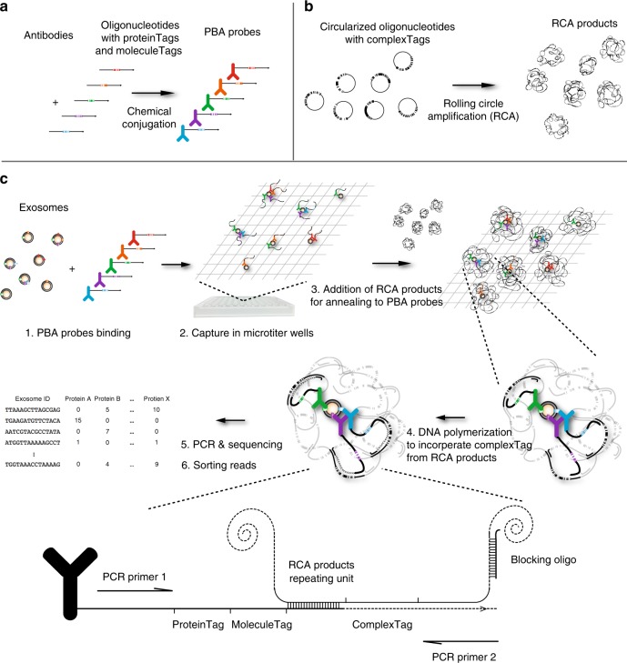
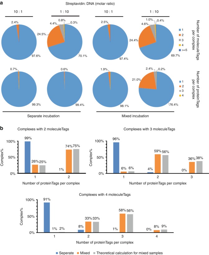
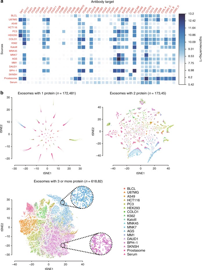
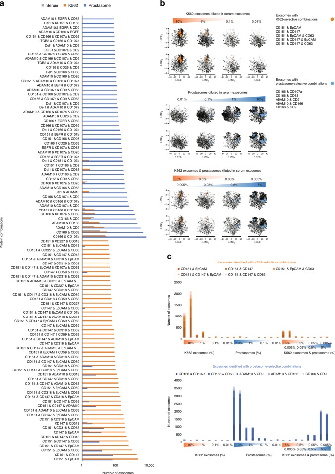
Exosomes have been implicated in numerous biological processes, and they may serve as important disease markers. Surface proteins on exosomes carry information about their tissues of origin. Because of the heterogeneity of exosomes it is desirable to investigate them individually, but this has so far remained impractical. Here, we demonstrate a proximity-dependent barcoding assay to profile surface proteins of individual exosomes using antibody-DNA conjugates and next-generation sequencing. We first validate the method using artificial streptavidin-oligonucleotide complexes, followed by analysis of the variable composition of surface proteins on individual exosomes, derived from human body fluids or cell culture media. Exosomes from different sources are characterized by the presence of specific combinations of surface proteins and their abundance, allowing exosomes to be separately quantified in mixed samples to serve as markers for tissue-specific engagement in disease.


Conflict of interest statement

D.W. has filed a patent application (PCT/SE2014/051133) describing the PBA technique. D.W., U.L., J.Y. and M.K-M hold shares in Vesicode AB commercializing the PBA technology. J.O. is an employee of Vesicode AB. The remaining authors declare no competing interests.

Figures

Fig. 1
Fig. 1
Design and workflow of PBA. a Preparation of PBA probes by chemical conjugation of antibodies and DNA oligonucleotides containing an 8-nt proteinTag and an 8-nt random sequence moleculeTag. b Preparation of RCA products from circularized oligonucleotides comprising a 15-nt random sequence as complexTags, capable of encoding in excess of one billion unique complexTags. c To profile surface proteins of exosomes by PBA, exosomes are first incubated with PBA probes, followed by capture of exosomes with bound PBA probes in microtiter wells via immobilized cholera toxin subunit B (CTB). Oligonucleotides on PBA probes brought together by binding the same exosome are next allowed to hybridize to a unique RCA product, followed by enzymatic extension, incorporating the complexTag present in the RCA product along with a standard sequence motif, later used for amplification. To prevent the DNA polymerase from extending across nearby monomers in the RCA products, blocking oligonucleotides are pre-hybridized to the RCA products. Successfully extended DNA molecules on PBA probes are amplified by PCR using the PCR primer1 and PCR primer2 for library preparation, while oligonucleotides on the antibodies or RCA products fail to be amplified by the PCR primer pairs used for amplification. The PCR product were subjected to DNA sequencing to record the numbers of molecules with specific tag combinations, thus revealing the identities of proteins on individual exosomes
Fig. 2
Fig. 2
Validation of PBA using STV-biotin-oligonucleotide complexes. Tetrameric streptavidin molecules were incubated with four different biotinylated oligonucleotides, either separately or in a mixture of all four oligonucleotides. Streptavidin and biotinylated oligonucleotides were combined at molar ratios of 1:10 or 10:1. a The numbers of observed complexes with different numbers of proteinTags and moleculeTags are summarized in pie charts. b When the oligonucleotides were in excess, the numbers of complexes from separate or mixed oligonucleotides incubation with 2, 3, or 4 moleculeTags were grouped according to the number of proteinTags per complex. Theoretical ratios were calculated for the mixed sample
Fig. 3
Fig. 3
Validation of PBA on exosomes using antibodies to CD9 and CD63. a Separate incubation: exosomes isolated from the K562 cell line were incubated with two distinct sets of PBA probes separately before they were pooled for PBA. b Mixed incubation: exosomes were incubated with four different PBA probes before PBA was performed
Fig. 4
Fig. 4
Bulk protein counts and visualization of individual exosomes. a, b Exosomes from 18 different samples were analyzed for the presence of 38 proteins (40 proteinTags) by PBA. a Heatmaps representing the total amounts of the proteins by log(moleculeTag + 1) found on exosomes from different sources. b Individual exosomes with one identified protein type, two protein types, and three or more protein types were visualized by t-SNE according to their protein compositions, with color and shape of the symbols representing the source of each exosome. Cells from two regions dominated by exosomes from the cell lines AGS and BPH-1 are highlighted in circles with dotted contours
Fig. 5
Fig. 5
Quantification of exosomes from different sources according to their surface protein combinations, as revealed by PBA. a Protein combinations selective for either K562 exosomes (orange) or prostasomes (blue), compared to exosomes from serum (gray), were sorted based on the number of exosomes with specific combinations. Here, the top 50 for each exosome are displayed. b, c Serial dilution of K562 exosomes or prostasomes spiked in serum exosomes were quantified using K562- or prostasomes-selective combinations, respectively. The exosomes identified with either K562- or prostasome-selective combinations are indicated by orange and blue filled circles in the tSNE plots in b, and with orange and blue bars, respectively, in the bar plots in c. The sizes of the colored circles in (b) are proportional to the number of exosomes with the same protein combination. Each two vertically adjacent tSNE plots and two horizontally adjacent bars are illustrating two replicates

References

    1. S ELA, Mager I, Breakefield XO, Wood MJ. Extracellular vesicles: biology and emerging therapeutic opportunities. Nat. Rev. Drug Discov. 2013;12:347–357. doi: 10.1038/nrd3978. - DOI - PubMed
    1. Hoshino A, et al. Tumour exosome integrins determine organotropic metastasis. Nature. 2015;527:329–335. doi: 10.1038/nature15756. - DOI - PMC - PubMed
    1. Costa-Silva B, et al. Pancreatic cancer exosomes initiate pre-metastatic niche formation in the liver. Nat. cell Biol. 2015;17:816–826. doi: 10.1038/ncb3169. - DOI - PMC - PubMed
    1. Yoshioka Y, et al. Ultra-sensitive liquid biopsy of circulating extracellular vesicles using ExoScreen. Nat. Commun. 2014;5:3591. doi: 10.1038/ncomms4591. - DOI - PMC - PubMed
    1. Santiago-Dieppa DR, et al. Extracellular vesicles as a platform for ‘liquid biopsy’ in glioblastoma patients. Expert Rev. Mol. Diagn. 2014;14:819–825. doi: 10.1586/14737159.2014.943193. - DOI - PMC - PubMed

Publication types

MeSH terms

LinkOut - more resources